

-
Product Name
Anti-Proteasome 20S LMP7 (3G3) Mouse antibody
- Documents
-
Description
Proteasome 20S LMP7 (3G3) Mouse monoclonal antibody
-
Tested applications
WB, IHC-P, ICC/IF
-
Species reactivity
Human, Rat
-
Isotype
Mouse IgG1
-
Preparation
Antigen: Purified recombinant fragment of human PSMB8 expressed in E. Coli.
-
Clonality
Monoclonal
-
Formulation
Ascitic fluid containing 0.03% sodium azide.
-
Storage instructions
Store at 4°C short term. Store at -20°C long term. Avoid freeze / thaw cycle.
-
Applications
WB: 1/500 - 1/2000
IHC: 1/200 - 1/1000
ICC: 1/200 - 1/1000
ELISA: 1/10000
-
Validations
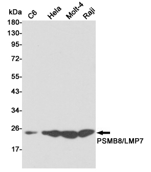
Western blot detection of PSMB8/LMP7 in C6,Hela,Molt4 and Raji cell lysates using PSMB8/LMP7 mouse mAb (1:1000 diluted).Predicted band size:30KDa.Observed band size:23KDa.
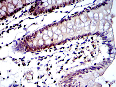
Immunohistochemical analysis of paraffin-embedded colon tissues using PSMB8 mouse mAb with DAB staining.
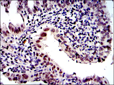
Immunohistochemical analysis of paraffin-embedded intima cancer tissues using PSMB8 mouse mAb with DAB staining.
Western blot analysis using PSMB8 mouse mAb against Hela (1), MCF-7 (2), A431 (3), RAJI (4), MOTL4 (5) and PC-12 (6) cell lysate.
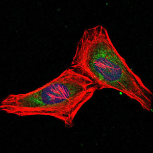
Immunofluorescence analysis of Hela cells using PSMB8 mouse mAb (green). Blue
-
Background
Swiss-Prot Acc.P28062.The proteasome is a multicatalytic proteinase complex with a highly ordered ring-shaped 20S core structure. The core structure is composed of 4 rings of 28 non-identical subunits; 2 rings are composed of 7 alpha subunits and 2 rings are composed of 7 beta subunits. Proteasomes are distributed throughout eukaryotic cells at a high concentration and cleave peptides in an ATP/ubiquitin-dependent process in a non-lysosomal pathway. An essential function of a modified proteasome, the immunoproteasome, is the processing of class I MHC peptides. This gene encodes a member of the proteasome B-type family, also known as the T1B family, that is a 20S core beta subunit. This gene is located in the class II region of the MHC (major histocompatibility complex). Expression of this gene is induced by gamma interferon and this gene product replaces catalytic subunit 3 (proteasome beta 5 subunit) in the immunoproteasome. Proteolytic processing is required to generate a mature subunit. Two alternative transcripts encoding two isoforms have been identified; both isoforms are processed to yield the same mature subunit.
Related Products / Services
Please note: All products are "FOR RESEARCH USE ONLY AND ARE NOT INTENDED FOR DIAGNOSTIC OR THERAPEUTIC USE"