
-
Product Name
Anti-SYNE1 antibody
- Documents
-
Description
Rabbit monoclonal antibody to SYNE1
-
Tested applications
WB, ICC, IF, IHC-P, FC
-
Species reactivity
Human, Mouse, Rat
-
Alternative names
8B antibody; AMC3 antibody; AMCM antibody; CPG2 antibody; ARCA1 antibody; EDMD4 antibody; KASH1 antibody; MYNE1 antibody; Nesp1 antibody; SCAR8 antibody; C6orf98 antibody; dJ45H2.2 antibody
-
Isotype
Rabbit IgG
-
Preparation
This antigen of this antibody was recombinant protein
-
Clonality
Monoclonal
-
Formulation
Liquid, 1*TBS (pH7.4), 0.05% BSA, 40% Glycerol. Preservative: 0.05% Sodium Azide.
-
Storage instructions
Store at +4℃ after thawing. Aliquot store at -20℃ or -80℃. Avoid repeated freeze / thaw cycles.
-
Applications
WB: 1:500-1:2,000
ICC: 1:200-1:1,000
IHC-P: 1:50-1:200
FC: 1:50-1:100
-
Validations
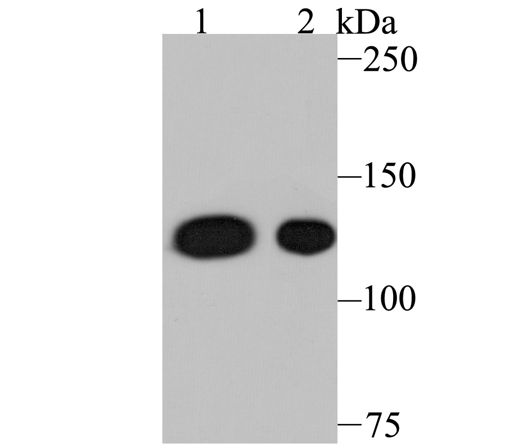
Fig1: Western blot analysis of Nesprin 1 on different lysates using anti-Nesprin 1 antibody at 1/500 dilution.; Positive control:; Lane 1: A549; Lane 2: Mouse spleen tissue
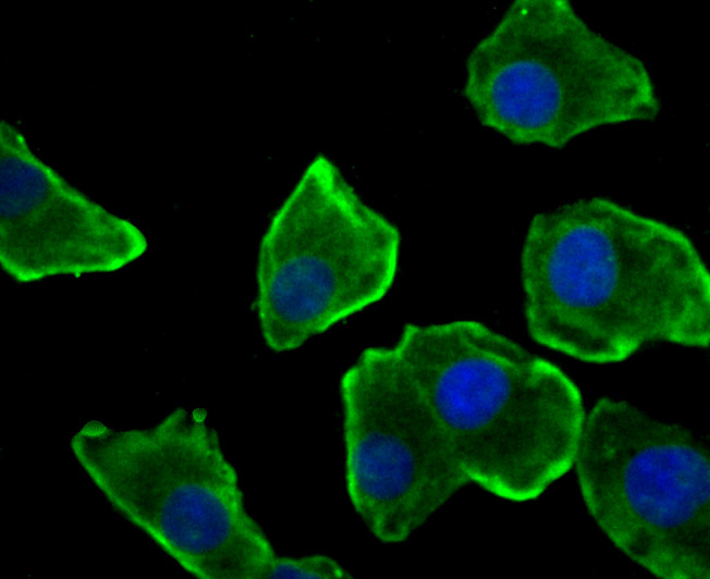
Fig2: ICC staining Nesprin 1 in A549 cells (green). The nuclear counter stain is DAPI (blue). Cells were fixed in paraformaldehyde, permeabilised with 0.25% Triton X100/PBS.
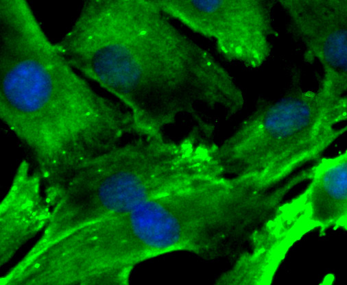
Fig3: ICC staining Nesprin 1 in C2C12 cells (green). The nuclear counter stain is DAPI (blue). Cells were fixed in paraformaldehyde, permeabilised with 0.25% Triton X100/PBS.
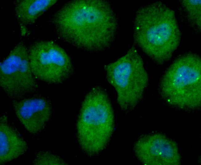
Fig4: ICC staining Nesprin 1 in HUVEC cells (green). The nuclear counter stain is DAPI (blue). Cells were fixed in paraformaldehyde, permeabilised with 0.25% Triton X100/PBS.
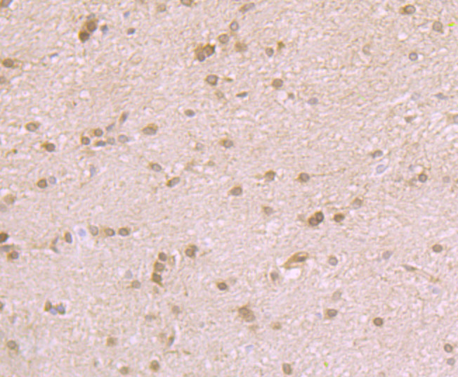
Fig5: Immunohistochemical analysis of paraffin-embedded rat brain tissue using anti-Nesprin 1 antibody. Counter stained with hematoxylin.
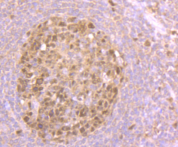
Fig6: Immunohistochemical analysis of paraffin-embedded human tonsil tissue using anti-Nesprin 1 antibody. Counter stained with hematoxylin.
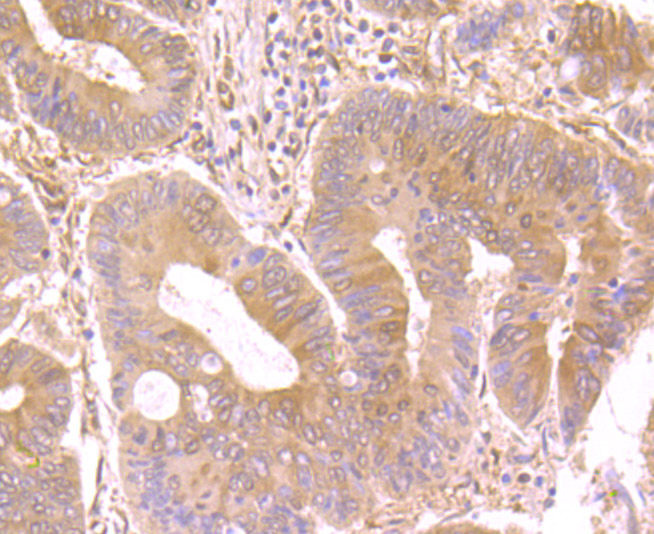
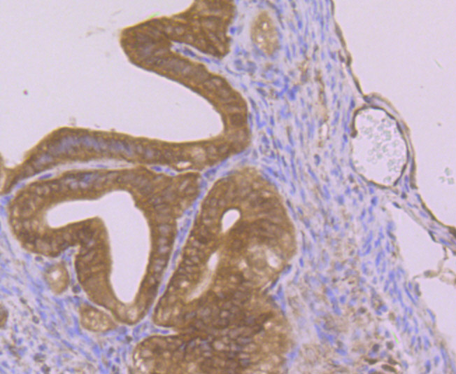
Fig7: Immunohistochemical analysis of paraffin-embedded human colon cancer tissue using anti-Nesprin 1 antibody. Counter stained with hematoxylin.
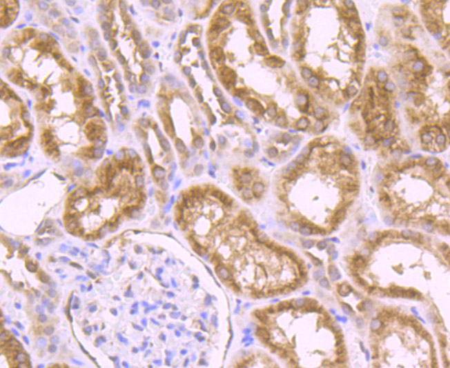
Fig8: Immunohistochemical analysis of paraffin-embedded human kidney tissue using anti-Nesprin 1 antibody. Counter stained with hematoxylin.
Fig9: Immunohistochemical analysis of paraffin-embedded mouse fallopian tubes tissue using anti-Nesprin 1 antibody. Counter stained with hematoxylin.
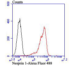
Fig10: Flow cytometric analysis of Daudi cells with Nesprin 1 antibody at 1/100 dilution (red) compared with an unlabelled control (cells without incubation with primary antibody; black).Alexa Fluor 488-conjugated goat anti-rabbit IgG was used as the seco
- Background
-
References
- Zhang Q et al. Nesprins: a novel family of spectrin-repeat-containing proteins that localize to the nuclear membrane in multiple tissues. J Cell Sci 114:4485-4498 (2001).
- Stewart-Hutchinson P J et al. Structural requirements for the assembly of LINC complexes and their function in cellular mechanical stiffness. Exp Cell Res 314: 1892-1905 (2008).
Related Products / Services
Please note: All products are "FOR RESEARCH USE ONLY AND ARE NOT INTENDED FOR DIAGNOSTIC OR THERAPEUTIC USE"