
-
Product Name
Anti-DYSF antibody
- Documents
-
Description
Rabbit polyclonal antibody to DYSF
-
Tested applications
WB, IHC-P, FC
-
Species reactivity
Human
-
Alternative names
MMD1 antibody; FER1L1 antibody; LGMD2B antibody; LGMDR2 antibody
-
Isotype
Rabbit IgG
-
Preparation
This antigen of this antibody was recombinant protein
-
Clonality
Polyclonal
-
Formulation
Liquid, 1*PBS (pH7.4), 0.2% BSA, 50% Glycerol. Preservative: 0.05% Sodium Azide.
-
Storage instructions
Store at +4℃ after thawing. Aliquot store at -20℃ or -80℃. Avoid repeated freeze / thaw cycles.
-
Applications
WB: 1:500-1:2,000
IHC-P: 1:50-1:200
FC: 1:50-1:100
-
Validations
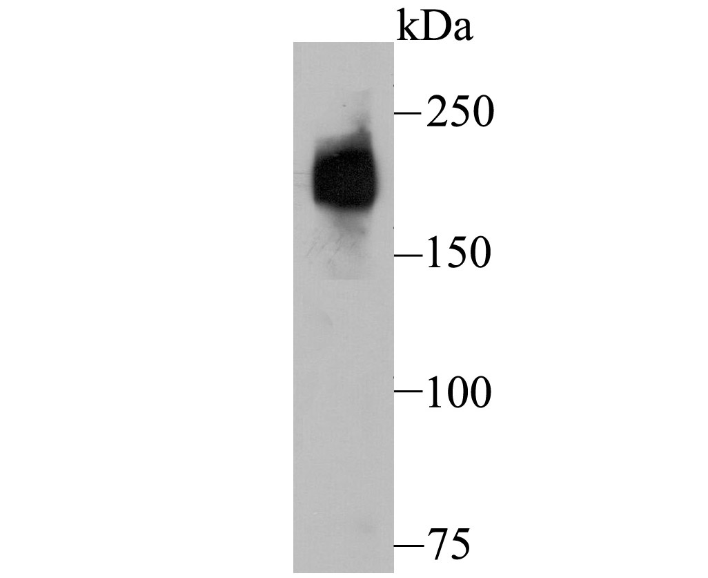
Fig1: Western blot analysis of Dysferlin on human fetal skeletal muscle tissue lysate using anti-Dysferlin antibody at 1/500 dilution.
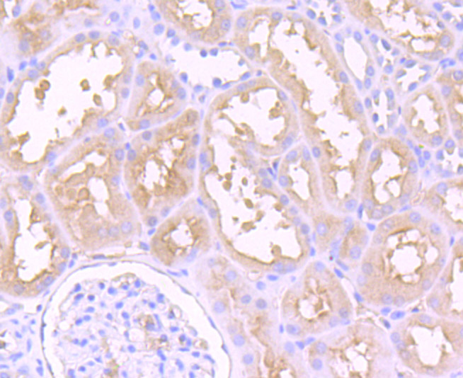
Fig2: Immunohistochemical analysis of paraffin-embedded human kidney tissue using anti-Dysferlin antibody. Counter stained with hematoxylin.
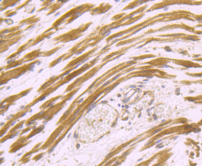
Fig3: Immunohistochemical analysis of paraffin-embedded human fetal skeletal muscle tissue using anti-Dysferlin antibody. Counter stained with hematoxylin.
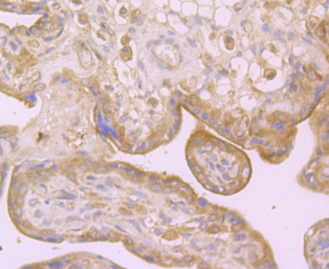
Fig4: Immunohistochemical analysis of paraffin-embedded human placenta tissue using anti-Dysferlin antibody. Counter stained with hematoxylin.
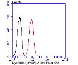
Fig5: Flow cytometric analysis of HUVEC cells with Dysferlin antibody at 1/100 dilution (red) compared with an unlabelled control (cells without incubation with primary antibody; black). Alexa Fluor 488-conjugated goat anti rabbit IgG was used as the secondary antibody.
- Background
-
References
- Fuson K et al. Alternate splicing of dysferlin C2A confers Ca(2+)-dependent and Ca(2+)-independent binding for membrane repair. Structure 22:104-115 (2014).
- Huang Y et al. AHNAK, a novel component of the dysferlin protein complex, redistributes to the cytoplasm with dysferlin during skeletal muscle regeneration. FASEB J 21:732-742 (2007).
Related Products / Services
Please note: All products are "FOR RESEARCH USE ONLY AND ARE NOT INTENDED FOR DIAGNOSTIC OR THERAPEUTIC USE"