
-
Product Name
UBE2T/HSPC150 antibody
- Documents
-
Description
UBE2T/HSPC150 Rabbit Polyclonal antibody. Positive IP detected in HeLa cells. Positive WB detected in Jurkat cells, HeLa cells, K-562 cells. Positive IF detected in HeLa cells, HepG2 cells. Observed molecular weight by Western-blot: 23 kDa
-
Tested applications
ELISA, WB, IP, IF
-
Species reactivity
Human,Mouse,Rat; other species not tested.
-
Alternative names
HSPC150 antibody; PIG50 antibody; UBE2T antibody; Ubiquitin carrier protein T antibody; Ubiquitin protein ligase T antibody
-
Isotype
Rabbit IgG
-
Preparation
This antibody was obtained by immunization of UBE2T/HSPC150 recombinant protein (Accession Number: NM_014176). Purification method: Antigen affinity purified.
-
Clonality
Polyclonal
-
Formulation
PBS with 0.1% sodium azide and 50% glycerol pH 7.3.
-
Storage instructions
Store at -20℃. DO NOT ALIQUOT
-
Applications
Recommended Dilution:
WB: 1:200-1:2000
IP: 1:200-1:2000
IF: 1:50-1:500
-
Validations
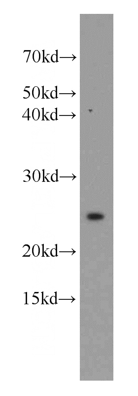
Jurkat cells were subjected to SDS PAGE followed by western blot with Catalog No:116537(UBE2T antibody) at dilution of 1:800
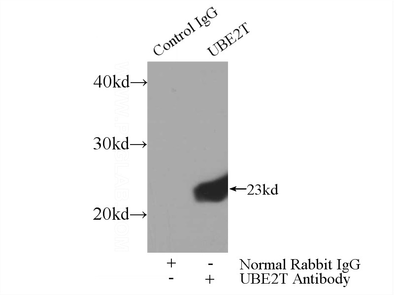
IP Result of anti-UBE2T (IP:Catalog No:116537, 3ug; Detection:Catalog No:116537 1:500) with HeLa cells lysate 3000ug.
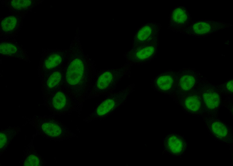
Immunofluorescent analysis of (10% Formaldehyde) fixed HeLa cells using Catalog No:116537(UBE2T Antibody) at dilution of 1:50 and Alexa Fluor 488-congugated AffiniPure Goat Anti-Rabbit IgG(H+L)
-
Background
The ubiquitin (Ub)-mediated protein degradation pathway involves three sequential enzymatic steps that facilitate the conjugation of Ub to specific protein substrates. The first step requires ATP-dependent activation of the C-terminus of Ub and the assembly of multi-Ubs by Ub-activating enzyme E1. The ubiquitin-conjugating enzyme E2, catalytic (UBCc) domain, is then conjugated to Ubs, through a thiol-ester linkage between a conserved cysteine and the C-terminus of Ub, to generate an intermediate Ub-E2 complex. Then the E3, a ligase, catalyzes the transfer of Ub from E2 to the appropriate substrate. This pathway regulates many fundamental cellular processes. There are also other E2s which form thiol-ester linkages without the use of E3s as well as several UBC homologs (TSG101, Mms2, Croc-1 and similar proteins), which lack the active site cysteine essential for ubiquitination and appear to function in DNA repair pathways.
-
References
- Hao J, Xu A, Xie X. Elevated expression of UBE2T in lung cancer tumors and cell lines. Tumour biology : the journal of the International Society for Oncodevelopmental Biology and Medicine. 29(3):195-203. 2008.
- Hira A, Yoshida K, Sato K. Mutations in the gene encoding the E2 conjugating enzyme UBE2T cause Fanconi anemia. American journal of human genetics. 96(6):1001-7. 2015.
- Jacquemont C, Taniguchi T. Proteasome function is required for DNA damage response and fanconi anemia pathway activation. Cancer research. 67(15):7395-405. 2007.
Related Products / Services
Please note: All products are "FOR RESEARCH USE ONLY AND ARE NOT INTENDED FOR DIAGNOSTIC OR THERAPEUTIC USE"