
-
Product Name
TNFR1 antibody
- Documents
-
Description
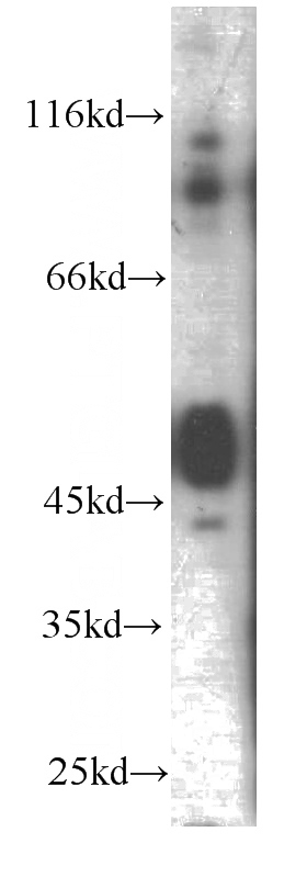
TNFR1 Mouse Monoclonal antibody. Positive WB detected in Human brain, HL-60 cells. Positive FC detected in Raji cells. Observed molecular weight by Western-blot: 50kd
-
Tested applications
ELISA, WB, FC
-
Species reactivity
Human,Mouse; other species not tested.
-
Alternative names
CD120a antibody; FPF antibody; p55 antibody; p55 R antibody; p60 antibody; TBP1 antibody; TBPI antibody; TNF R antibody; TNF R I antibody; TNF R1 antibody; TNF R55 antibody; TNF Receptor 1 antibody; TNF Receptor I antibody; TNF RI antibody; TNFAR antibody; TNFR I antibody; TNFR1 antibody; TNFR55 antibody; TNFR60 antibody; TNFRSF1A antibody
-
Isotype
Mouse IgG1
-
Preparation
This antibody was obtained by immunization of TNFR1 recombinant protein (Accession Number: NM_001065). Purification method: Protein G purified.
-
Clonality
Monoclonal
-
Formulation
PBS with 0.02% sodium azide and 50% glycerol pH 7.3.
-
Storage instructions
Store at -20℃. DO NOT ALIQUOT
-
Applications
Recommended Dilution:
WB: 1:500-1:5000
-
Validations
human brain tissue were subjected to SDS PAGE followed by western blot with Catalog No:107631(TNFR1 antibody) at dilution of 1:1000
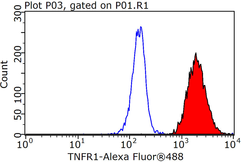
1X10^6 Raji cells were stained with 0.2ug TNFR1 antibody (Catalog No:107631, red) and control antibody (blue). Fixed with 90% MeOH blocked with 3% BSA (30 min). Alexa Fluor 488-congugated AffiniPure Goat Anti-Mouse IgG(H+L) with dilution 1:1000.
-
Background
Tumor necrosis factor (TNF) is a multifunctional cytokine that plays a key role in regulating inflammation, immune functions, host defense, and apoptosis (PMID: 16407280). TNF exists in soluble and membrane-bound forms. TNF signals through two distinct cell surface receptors, TNFR1 (TNFRSF1A, CD120a) and TNFR2 (TNFRSF1B, CD120b). Whereas TNFR1 is widely expressed, expression of TNFR2 is limited to cells of the immune system, endothelial cells, and nerve cells (PMID: 22053109). TNFR1, which contains a death domain (DD) within its intracytoplasmic region, is thought to be the key receptor for TNF signaling (PMID: 16407280). This receptor can activate NF-kappaB, mediate apoptosis, and function as a regulator of inflammation. Antiapoptotic protein BCL2-associated athanogene 4 (BAG4/SODD) and adaptor proteins TRADD and TRAF2 have been shown to interact with this receptor, and thus play regulatory roles in the signal transduction mediated by the receptor.
Related Products / Services
Please note: All products are "FOR RESEARCH USE ONLY AND ARE NOT INTENDED FOR DIAGNOSTIC OR THERAPEUTIC USE"