
-
Product Name
TDP-43 antibody
- Documents
-
Description
TDP-43 Rabbit Polyclonal antibody. Positive WB detected in K-562 cells, A549 cells, HeLa cells, HL-60 cells, human heart tissue, mouse brain tissue, mouse pancreas tissue, rat brain tissue. Positive IP detected in HeLa cells, K-562 cells. Positive FC detected in HeLa cells. Positive IF detected in HeLa cells, HepG2 cells, MCF-7 cells, SH-SY5Y cells. Positive IHC detected in human brain (FTLD-U) tissue, human brain tissue, human gliomas tissue, human pancreas tissue, human prostate cancer tissue, mouse brain tissue, rat brain tissue. Observed molecular weight by Western-blot: 43 kDa
-
Tested applications
ELISA, WB, IHC, IP, FC, IF
-
Species reactivity
Human,Mouse,Rat,Zebrafish; other species not tested.
-
Alternative names
ALS10 antibody; TAR DNA binding protein antibody; TAR DNA binding protein 43 antibody; TARDBP antibody; TDP 43 antibody; TDP43 antibody
-
Isotype
Rabbit IgG
-
Preparation
This antibody was obtained by immunization of TDP-43 recombinant protein (Accession Number: BC001487). Purification method: Antigen affinity purified.
-
Clonality
Polyclonal
-
Formulation
PBS with 0.1% sodium azide and 50% glycerol pH 7.3.
-
Storage instructions
Store at -20℃. DO NOT ALIQUOT
-
Applications
Recommended Dilution:
WB: 1:1000-1:10000
IP: 1:200-1:2000
IHC: 1:20-1:200
IF: 1:20-1:200
-
Validations
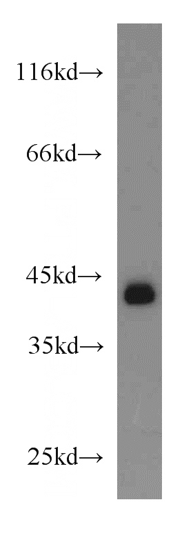
K-562 cells were subjected to SDS PAGE followed by western blot with Catalog No:115925(TARDBP antibody) at dilution of 1:2000
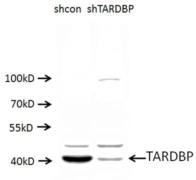
A549 cells (shcontrol and shRNA of TDP43) were subjected to SDS PAGE followed by western blot with Catalog No:115925 (TARDBP antibody) at dilution of 1:1000. (Data provided by Angran Biotech (www.miRNAlab.com)).
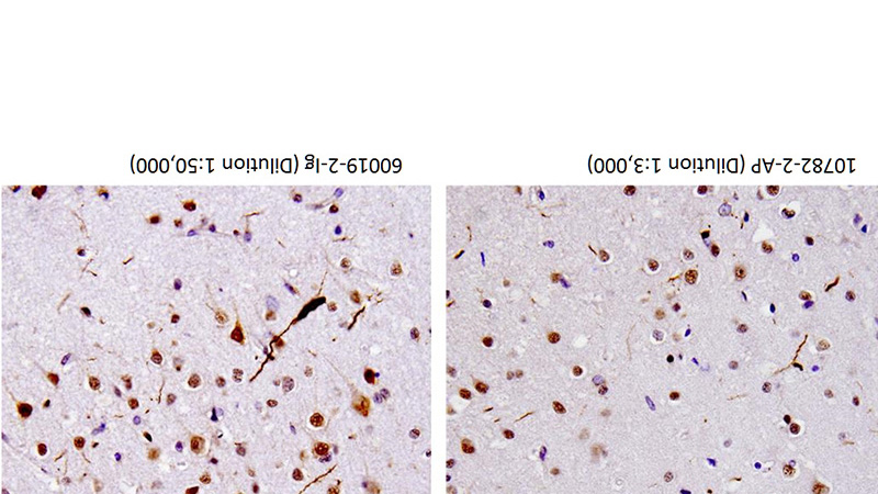
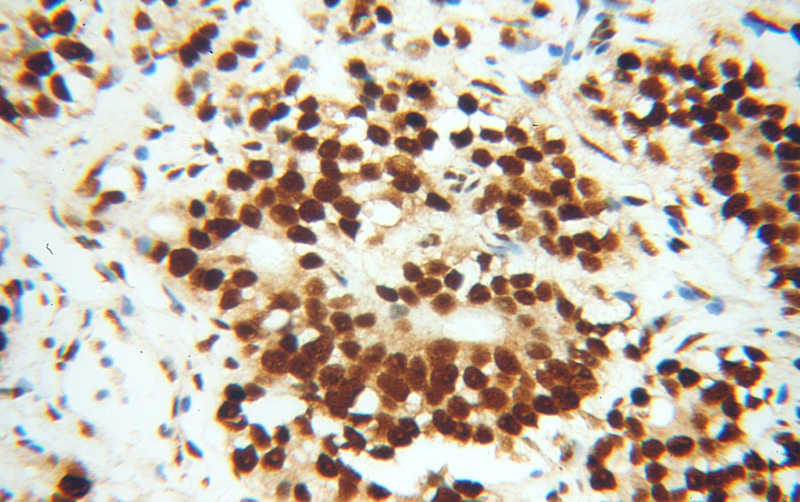
40X of FTLD-U case stained by Catalog No:115925 and Catalog No:107618, showing dystrophic neurites. (Figs were provided by Linda K. Kwong)
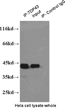
IP result of anti-TDP43(Catalog No:115925 for IP and Catalog No:107618 for Detection).
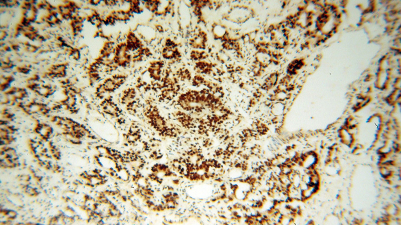
Immunohistochemical of paraffin-embedded human gliomas using Catalog No:115925(TARDBP antibody) at dilution of 1:200 (under 40x lens)
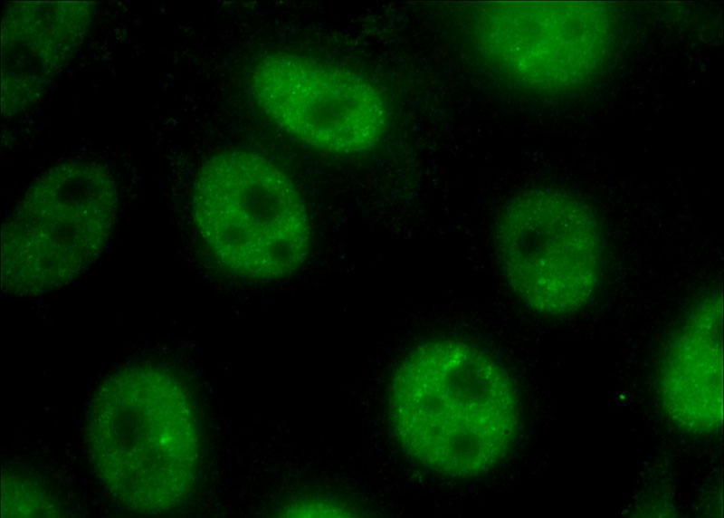
Immunofluorescent analysis of (10% Formaldehyde) fixed HeLa cells using Catalog No:115925(TDP-43 Antibody) at dilution of 1:400 and Alexa Fluor 488-congugated AffiniPure Goat Anti-Rabbit IgG(H+L)
Immunohistochemical of paraffin-embedded human gliomas using Catalog No:115925(TARDBP antibody) at dilution of 1:200 (under 10x lens)
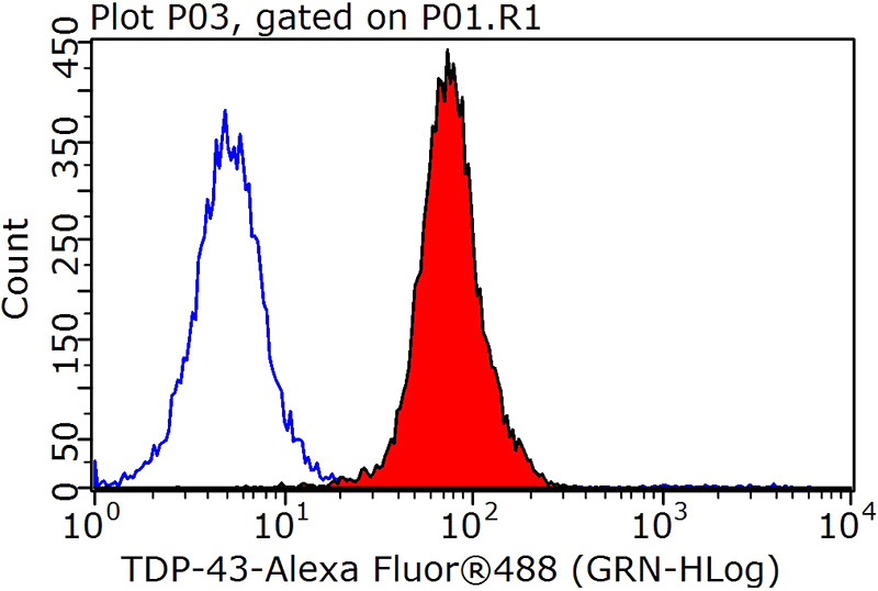
1X10^6 HeLa cells were stained with 0.2ug TDP-43 antibody (Catalog No:115925, red) and control antibody (blue). Fixed with 90% MeOH blocked with 3% BSA (30 min). Alexa Fluor 488-congugated AffiniPure Goat Anti-Rabbit IgG(H+L) with dilution 1:1000.
-
Background
The TARDBP gene encodes the TDP-43 protein, initially found to repress HIV-1 transcription by binding TAR DNA. TDP-43 has since been shown to bind RNA as well as DNA, and have multiple functions in transcriptional repression, translational regulation and pre-mRNA splicing. For instance, it is reported to regulate alternate splicing of the CTFR gene. In 2006 Neumann et al. found that hyperphosphorylated, ubiquitinated and/or cleaved forms of TDP-43, collectively known as pathological TDP-43, play a major role in the disease mechanisms of ubiquitin-positive, tau- and alpha-synuclein-negative frontotemporal dementia (FTLD-U) and in amyotrophic lateral sclerosis (ALS). Proteintech’s 10782-2-AP antibody is a rabbit polyclonal antibody recognizing N-terminal TDP-43. Generated using the first 260 amino acids of TDP-43, it recognizes the intact 45 kDa protein as well as all posttranslationally modified and truncated forms in multiple applications..
-
References
- Zhang T, Baldie G, Periz G, Wang J. RNA-processing protein TDP-43 regulates FOXO-dependent protein quality control in stress response. PLoS genetics. 10(10):e1004693. 2014.
- Kim KY, Lee HW, Shim YM, Mook-Jung I, Jeon GS, Sung JJ. A phosphomimetic mutant TDP-43 (S409/410E) induces Drosha instability and cytotoxicity in Neuro 2A cells. Biochemical and biophysical research communications. 464(1):236-43. 2015.
- Ward ME, Taubes A, Chen R. Early retinal neurodegeneration and impaired Ran-mediated nuclear import of TDP-43 in progranulin-deficient FTLD. The Journal of experimental medicine. 211(10):1937-45. 2014.
- Colombrita C, Onesto E, Buratti E. From transcriptomic to protein level changes in TDP-43 and FUS loss-of-function cell models. Biochimica et biophysica acta. 1849(12):1398-410. 2015.
- Xia Q, Wang H, Hao Z. TDP-43 loss of function increases TFEB activity and blocks autophagosome-lysosome fusion. The EMBO journal. 35(2):121-42. 2016.
- Wilson-Edell KA, Kehasse A, Scott GK. RPL24: a potential therapeutic target whose depletion or acetylation inhibits polysome assembly and cancer cell growth. Oncotarget. 5(13):5165-76. 2014.
- Zhan L, Xie Q, Tibbetts RS. Opposing roles of p38 and JNK in a Drosophila model of TDP-43 proteinopathy reveal oxidative stress and innate immunity as pathogenic components of neurodegeneration. Human molecular genetics. 24(3):757-72. 2015.
- Nandar W, Neely EB, Simmons Z, Connor JR. H63D HFE genotype accelerates disease progression in animal models of amyotrophic lateral sclerosis. Biochimica et biophysica acta. 1842(12 Pt A):2413-26. 2014.
Related Products / Services
Please note: All products are "FOR RESEARCH USE ONLY AND ARE NOT INTENDED FOR DIAGNOSTIC OR THERAPEUTIC USE"