
-
Product Name
TBR1 antibody
- Documents
-
Description
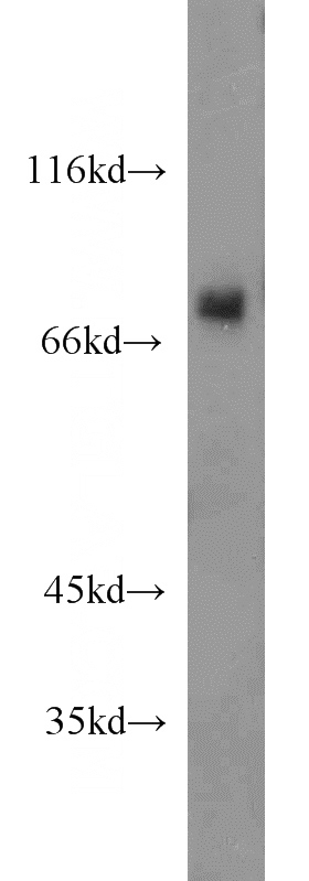
TBR1 Rabbit Polyclonal antibody. Positive IP detected in mouse brain tissue. Positive WB detected in mouse brain tissue. Positive IHC detected in human brain tissue. Observed molecular weight by Western-blot: 74kd
-
Tested applications
ELISA, WB, IHC, IP
-
Species reactivity
Human,Mouse,Rat; other species not tested.
-
Alternative names
T box brain protein 1 antibody; T box antibody; brain antibody; T brain 1 antibody; TBR 1 antibody; TBR1 antibody; TES 56 antibody
-
Isotype
Rabbit IgG
-
Preparation
This antibody was obtained by immunization of TBR1 recombinant protein (Accession Number: NM_006593). Purification method: Antigen affinity purified.
-
Clonality
Polyclonal
-
Formulation
PBS with 0.02% sodium azide and 50% glycerol pH 7.3.
-
Storage instructions
Store at -20℃. DO NOT ALIQUOT
-
Applications
Recommended Dilution:
WB: 1:500-1:5000
IP: 1:200-1:2000
IHC: 1:20-1:200
-
Validations
mouse brain tissue were subjected to SDS PAGE followed by western blot with Catalog No:115881(TBR1 antibody) at dilution of 1:1500
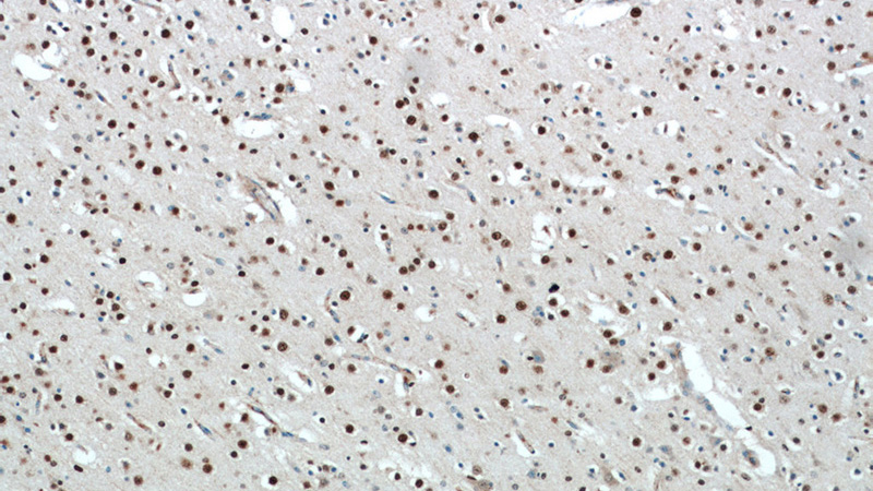
Immunohistochemical of paraffin-embedded human brain using Catalog No:115881(TBR1 antibody) at dilution of 1:100 (under 10x lens)
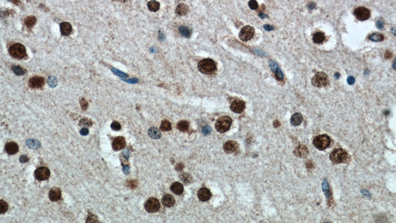
Immunohistochemical of paraffin-embedded human brain using Catalog No:115881(TBR1 antibody) at dilution of 1:100 (under 40x lens)
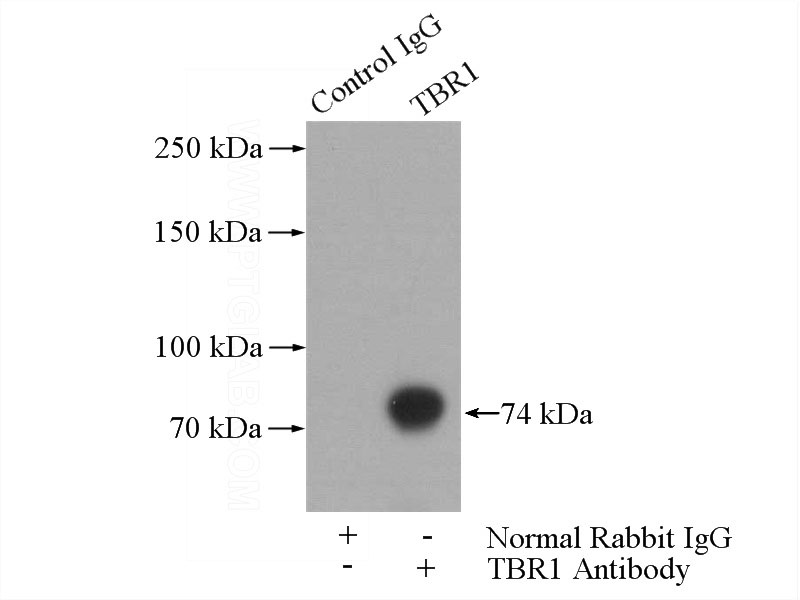
IP Result of anti-TBR1 (IP:Catalog No:115881, 4ug; Detection:Catalog No:115881 1:600) with mouse brain tissue lysate 4000ug.
-
Background
TBR1, also named as T-box brain protein 1, is a 682 amino acid protein, which contains one T-box DNA-binding domain and localizes in the nucleus. TBR1 is expressed in the brain and as a transcriptional regulator is involved in developmental processes. TBR1 is required for normal brain development.
-
References
- Denton KR, Lei L, Grenier J, Rodionov V, Blackstone C, Li XJ. Loss of spastin function results in disease-specific axonal defects in human pluripotent stem cell-based models of hereditary spastic paraplegia. Stem cells (Dayton, Ohio). 32(2):414-23. 2014.
- Zhu PP, Denton KR, Pierson TM, Li XJ, Blackstone C. Pharmacologic rescue of axon growth defects in a human iPSC model of hereditary spastic paraplegia SPG3A. Human molecular genetics. 23(21):5638-48. 2014.
- Radonjić NV, Memi F, Ortega JA, Glidden N, Zhan H, Zecevic N. The Role of Sonic Hedgehog in the Specification of Human Cortical Progenitors In Vitro. Cerebral cortex (New York, N.Y. : 1991). 26(1):131-43. 2016.
- Radonjić NV, Ortega JA, Memi F, Dionne K, Jakovcevski I, Zecevic N. The complexity of the calretinin-expressing progenitors in the human cerebral cortex. Frontiers in neuroanatomy. 8:82. 2014.
- Denton KR, Xu CC, Li XJ. Modeling Axonal Phenotypes with Human Pluripotent Stem Cells. Methods in molecular biology (Clifton, N.J.). 1353:309-21. 2016.
- Pan X, Chang X, Leung C. PAK1 regulates cortical development via promoting neuronal migration and progenitor cell proliferation. Molecular brain. 8:36. 2015.
- Radonjić NV, Ayoub AE, Memi F. Diversity of cortical interneurons in primates: the role of the dorsal proliferative niche. Cell reports. 9(6):2139-51. 2014.
- Xu CC, Denton KR, Wang ZB, Zhang X, Li XJ. Abnormal mitochondrial transport and morphology as early pathological changes in human models of spinal muscular atrophy. Disease models & mechanisms. 9(1):39-49. 2016.
Related Products / Services
Please note: All products are "FOR RESEARCH USE ONLY AND ARE NOT INTENDED FOR DIAGNOSTIC OR THERAPEUTIC USE"