
-
Product Name
TAB2 antibody
- Documents
-
Description
TAB2 Rabbit Polyclonal antibody. Positive WB detected in HepG2 cells, human liver tissue, L02 cells. Positive IP detected in HepG2 cells. Positive IF detected in HepG2 cells. Positive IHC detected in human malignant melanoma tissue. Positive FC detected in HepG2 cells. Observed molecular weight by Western-blot: 77 kDa
-
Tested applications
ELISA, IHC, IF, WB, FC, IP
-
Species reactivity
Human,Mouse,Rat; other species not tested.
-
Alternative names
FLJ21885 antibody; KIAA0733 antibody; MAP3K7IP2 antibody; TAB 2 antibody; TAB2 antibody; TAK1 binding protein 2 antibody
-
Isotype
Rabbit IgG
-
Preparation
This antibody was obtained by immunization of TAB2 recombinant protein (Accession Number: XM_047418490). Purification method: Antigen affinity purified.
-
Clonality
Polyclonal
-
Formulation
PBS with 0.02% sodium azide and 50% glycerol pH 7.3.
-
Storage instructions
Store at -20℃. DO NOT ALIQUOT
-
Applications
Recommended Dilution:
WB: 1:200-1:2000
IP: 1:200-1:2000
IHC: 1:20-1:200
IF: 1:20-1:200
-
Validations
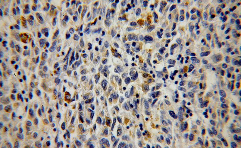
Immunohistochemical of paraffin-embedded human malignant melanoma using Catalog No:115965(MAP3K7IP2 antibody) at dilution of 1:100 (under 40x lens)
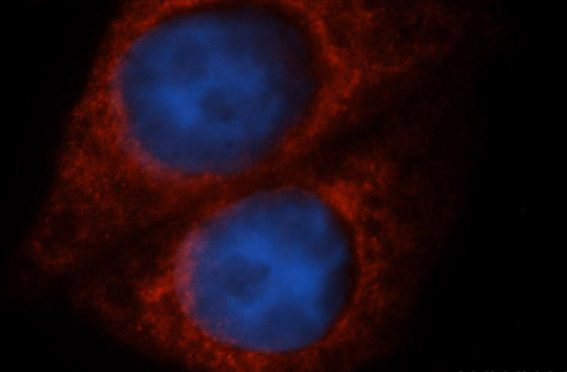
Immunofluorescent analysis of HepG2 cells, using MAP3K7IP2 antibody Catalog No:115965 at 1:50 dilution and Rhodamine-labeled goat anti-rabbit IgG (red). Blue pseudocolor = DAPI (fluorescent DNA dye).
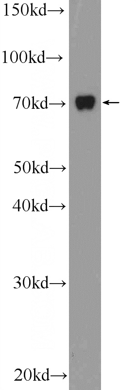
HepG2 cells were subjected to SDS PAGE followed by western blot with Catalog No:115965(MAP3K7IP2 Antibody) at dilution of 1:600
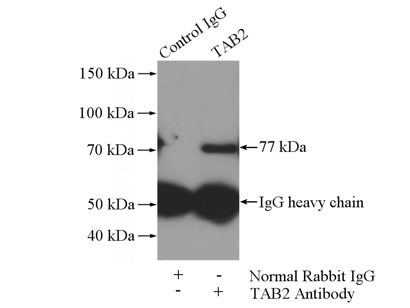
IP Result of anti-MAP3K7IP2 (IP:Catalog No:115965, 4ug; Detection:Catalog No:115965 1:600) with HepG2 cells lysate 3600ug.
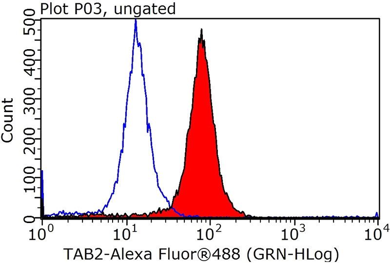
1X10^6 HepG2 cells were stained with 0.2ug MAP3K7IP2 antibody (Catalog No:115965, red) and control antibody (blue). Fixed with 90% MeOH blocked with 3% BSA (30 min). Alexa Fluor 488-congugated AffiniPure Goat Anti-Rabbit IgG(H+L) with dilution 1:1000.
-
Background
Tab2 has a role in the inflammatory signal transduction pathway and is involved in the reversal of NCoR-dependent and NFkB- or APP-mediated gene repression and androgen antagonist response. Besides, it is identified as a cofactor for ERBB4 to assemble NCoR corepressor complex in response to neuregulin [PMID:16469706]. It also functions as an adaptor that links TAK1 and TRAF6 in response to interleukin-1β (IL-1β) or tumour necrosis factor-α (TNF-α), and thereby mediates TAK1 activation [PMID:11460167].
Related Products / Services
Please note: All products are "FOR RESEARCH USE ONLY AND ARE NOT INTENDED FOR DIAGNOSTIC OR THERAPEUTIC USE"