
-
Product Name
STAU1 antibody
- Documents
-
Description
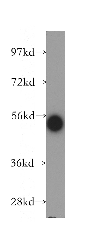
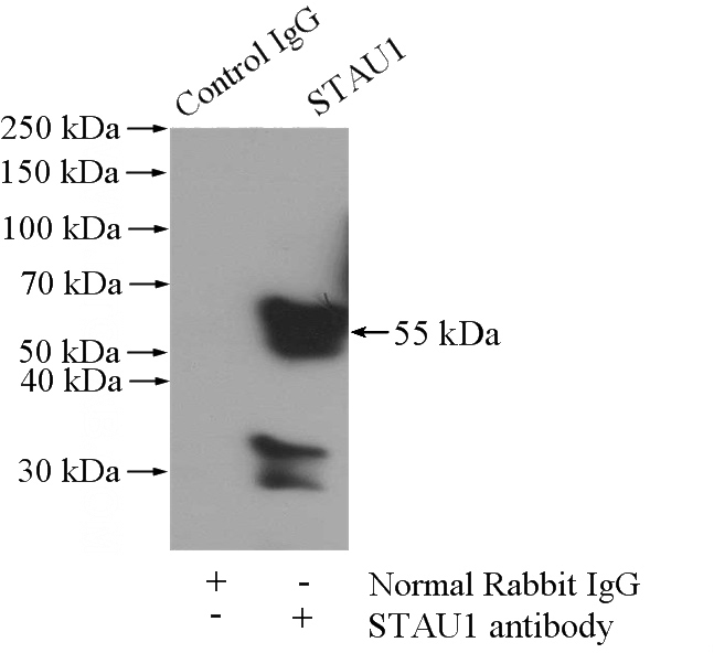
STAU1 Rabbit Polyclonal antibody. Positive IP detected in mouse brain tissue. Positive WB detected in human brain tissue, K-562 cells, mouse brain tissue. Observed molecular weight by Western-blot: 55 kDa
-
Tested applications
ELISA, WB, IP
-
Species reactivity
Human,Mouse,Rat; other species not tested.
-
Alternative names
FLJ25010 antibody; STAU antibody; STAU1 antibody
-
Isotype
Rabbit IgG
-
Preparation
This antibody was obtained by immunization of STAU1 recombinant protein (Accession Number: NM_001322930). Purification method: Antigen affinity purified.
-
Clonality
Polyclonal
-
Formulation
PBS with 0.02% sodium azide and 50% glycerol pH 7.3.
-
Storage instructions
Store at -20℃. DO NOT ALIQUOT
-
Applications
Recommended Dilution:
WB: 1:500-1:5000
IP: 1:200-1:2000
-
Validations
human brain tissue were subjected to SDS PAGE followed by western blot with Catalog No:115699(STAU1 antibody) at dilution of 1:300
IP Result of anti-STAU1 (IP:Catalog No:115699, 4ug; Detection:Catalog No:115699 1:500) with mouse brain tissue lysate 3440ug.
-
Background
Staufen1 (STAU1)-mediated mRNA decay (SMD) degrades translationally active mRNAs that bind the double-stranded (ds)RNA binding protein STAU1 within their 3'-untranslated regions (3'UTRs) [PMID:21307942]. STAU1 can binds to stem structures and protect Dvl2 mRNA from degradation by associating with the secondary structure in the U1 region which is located at 5′ position of second U stretch in the U2 region [PMID:17510634]. It has a role in specific positioning of mRNAs at given sites in the cell by cross-linking cytoskeletal and RNA components, and in stimulating their translation at the site.
-
References
- Sugimoto Y, Vigilante A, Darbo E. hiCLIP reveals the in vivo atlas of mRNA secondary structures recognized by Staufen 1. Nature. 519(7544):491-4. 2015.
- Alami NH, Smith RB, Carrasco MA. Axonal transport of TDP-43 mRNA granules is impaired by ALS-causing mutations. Neuron. 81(3):536-43. 2014.
Related Products / Services
Please note: All products are "FOR RESEARCH USE ONLY AND ARE NOT INTENDED FOR DIAGNOSTIC OR THERAPEUTIC USE"