
-
Product Name
SMAD2 antibody
- Documents
-
Description
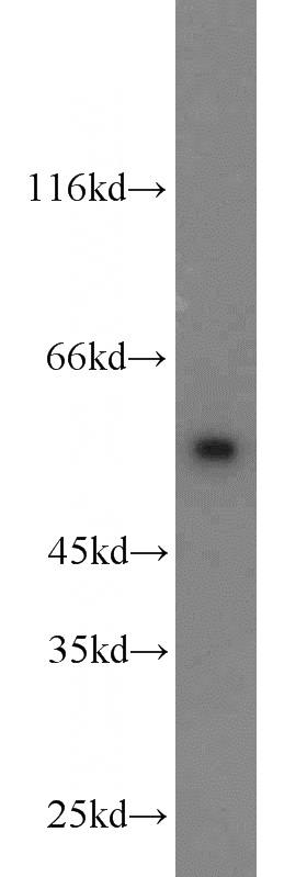
SMAD2 Rabbit Polyclonal antibody. Positive IP detected in HepG2 cells. Positive WB detected in HeLa cells, HepG2 cells, Jurkat cells, K-562 cells, mouse skeletal muscle tissue, rat skeletal muscle tissue. Positive IF detected in HepG2 cells. Positive IHC detected in human endometrial cancer tissue, human cervical cancer tissue, human heart tissue. Observed molecular weight by Western-blot: 58kd
-
Tested applications
ELISA, WB, IHC, IF, IP
-
Species reactivity
Human,Mouse,Rat; other species not tested.
-
Alternative names
hMAD 2 antibody; hSMAD2 antibody; JV18 antibody; JV18 1 antibody; MAD homolog 2 antibody; Mad related protein 2 antibody; MADH2 antibody; MADR2 antibody; Mothers against DPP homolog 2 antibody; SMAD 2 antibody; SMAD family member 2 antibody; SMAD2 antibody
-
Isotype
Rabbit IgG
-
Preparation
This antibody was obtained by immunization of SMAD2 recombinant protein (Accession Number: XM_006722451). Purification method: Antigen affinity purified.
-
Clonality
Polyclonal
-
Formulation
PBS with 0.02% sodium azide and 50% glycerol pH 7.3.
-
Storage instructions
Store at -20℃. DO NOT ALIQUOT
-
Applications
Recommended Dilution:
WB: 1:500-1:5000
IP: 1:500-1:5000
IHC: 1:20-1:200
IF: 1:10-1:100
-
Validations
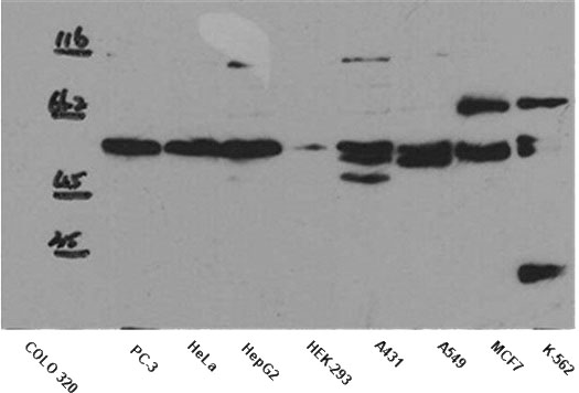
WB result of anti-SMAD2 (Catalog No:115416) in different cell lysates.
HeLa cells were subjected to SDS PAGE followed by western blot with Catalog No:115416(SMAD2 antibody) at dilution of 1:1000
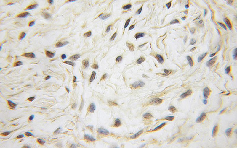
Immunohistochemical of paraffin-embedded human endometrial cancer using Catalog No:115416(SMAD2 antibody) at dilution of 1:50 (under 10x lens)
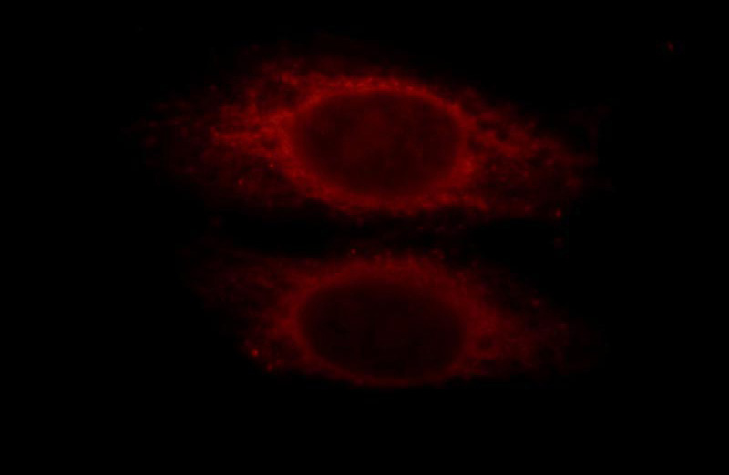
Immunofluorescent analysis of HepG2 cells, using SMAD2 antibody Catalog No:115416 at 1:25 dilution and Rhodamine-labeled goat anti-rabbit IgG (red).
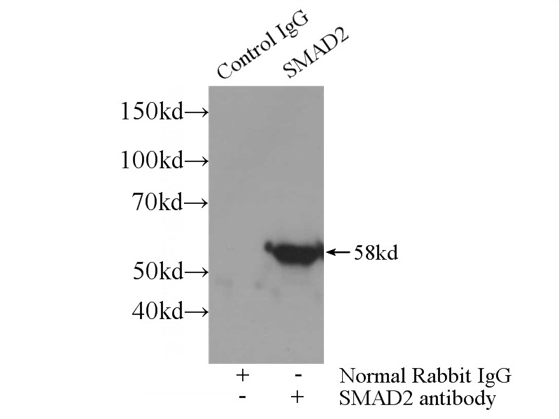
IP Result of anti-SMAD2 (IP:Catalog No:115416, 3ug; Detection:Catalog No:115416 1:1000) with HepG2 cells lysate 3000ug.
-
Background
SMAD2, also named as MADH2 and MADR2, belongs to the dwarfin/SMAD family, contains 1 MH1 (MAD homology 1) domain and 1 MH2 (MAD homology 2) domain. SMAD2 is a receptor-regulated SMAD(R-SMAD) that is an intracellular signal transducer and transcriptional modulator activated by TGF-beta (transforming growth factor) and activin type 1 receptor kinases. This protein may act as a tumor suppressor in colorectal carcinoma. It is phosphorylated on one or several of Thr-220, Ser-245, Ser-250, and Ser-255. In response to TGF-beta, It is phosphorylated on Ser-465/467 by TGF-beta and activin type 1 receptor kinases, and then able to interact with SMURF2, recruiting other proteins, such as SNON, for degradation. In response to decorin, the naturally occurring inhibitor of TGF-beta signaling, it is phosphorylated on Ser-240 by CaMK2. It is phosphorylated by MAPK3 upon EGF stimulation; which increases transcriptional activity and stability, and is blocked by calmodulin. In response to TGF-beta, it is ubiquitinated by NEDD4L, which promotes its degradation. In response to TGF-beta signaling, it is acetylated on Lys-19 by coactivators, which increases transcriptional activity. This antibody is a rabbit polyclonal antibody raised against residues near the N terminus of human SMAD2.
-
References
- Ma B, Kang Q, Qin L, Cui L, Pei C. TGF-β2 induces transdifferentiation and fibrosis in human lens epithelial cells via regulating gremlin and CTGF. Biochemical and biophysical research communications. 447(4):689-95. 2014.
- Shen M, Chen K, Lu J. Protective effect of astaxanthin on liver fibrosis through modulation of TGF-β1 expression and autophagy. Mediators of inflammation. 2014:954502. 2014.
- Chen MS, Zhang JH, Wang JL, Gao L, Chen XX, Xiao JH. Anti-fibrotic effects of neferine on carbon tetrachloride-induced hepatic fibrosis in mice. The American journal of Chinese medicine. 43(2):231-40. 2015.
- Dong D, Yin L, Qi Y, Xu L, Peng J. Protective Effect of the Total Saponins from Rosa laevigata Michx Fruit against Carbon Tetrachloride-Induced Liver Fibrosis in Rats. Nutrients. 7(6):4829-50. 2015.
- Zhang X, Han X, Yin L. Potent effects of dioscin against liver fibrosis. Scientific reports. 5:9713. 2015.
- Zhang X, Xiao J, Li R. Metformin alleviates vascular calcification induced by vitamin D3 plus nicotine in rats via the AMPK pathway. Vascular pharmacology. 2016.
- You SP, Ma L, Zhao J, Zhang SL, Liu T. Phenylethanol Glycosides from Cistanche tubulosa Suppress Hepatic Stellate Cell Activation and Block the Conduction of Signaling Pathways in TGF-β₁/smad as Potential Anti-Hepatic Fibrosis Agents. Molecules (Basel, Switzerland). 21(1):. 2016.
- Zhang Y, Fei M, Xue G. Elevated levels of hypoxia-inducible microRNA-210 in pre-eclampsia: new insights into molecular mechanisms for the disease. Journal of cellular and molecular medicine. 16(2):249-59. 2012.
Related Products / Services
Please note: All products are "FOR RESEARCH USE ONLY AND ARE NOT INTENDED FOR DIAGNOSTIC OR THERAPEUTIC USE"