
-
Product Name
SHARPIN antibody
- Documents
-
Description
SHARPIN Rabbit Polyclonal antibody. Positive WB detected in Raji cells, A549 cells, Jurkat cells. Positive IP detected in Raji cells. Positive IHC detected in human small intestine tissue. Observed molecular weight by Western-blot: 40kd
-
Tested applications
ELISA, WB, IP, IHC
-
Species reactivity
Human,Mouse,Rat; other species not tested.
-
Alternative names
DKFZp434N1923 antibody; hSIPL1 antibody; SHARPIN antibody; SIPL1 antibody
-
Isotype
Rabbit IgG
-
Preparation
This antibody was obtained by immunization of SHARPIN recombinant protein (Accession Number: XM_017013888). Purification method: Antigen affinity purified.
-
Clonality
Polyclonal
-
Formulation
PBS with 0.02% sodium azide and 50% glycerol pH 7.3.
-
Storage instructions
Store at -20℃. DO NOT ALIQUOT
-
Applications
Recommended Dilution:
WB: 1:500-1:5000
IP: 1:500-1:5000
IHC: 1:20-1:200
-
Validations
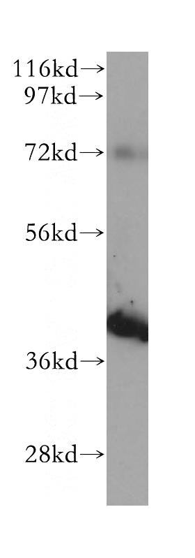
Raji cells were subjected to SDS PAGE followed by western blot with Catalog No:115264(SHARPIN antibody) at dilution of 1:500
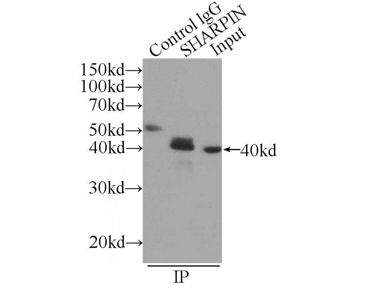
IP Result of anti-SHARPIN (IP:Catalog No:115264, 3ug; Detection:Catalog No:115264 1:1500) with Raji cells lysate 2520ug.
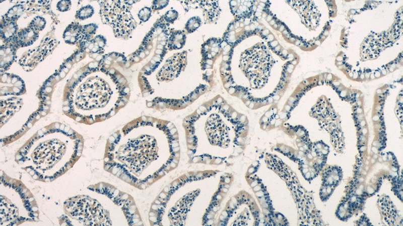
Immunohistochemistry of paraffin-embedded human small intestine tissue slide using Catalog No:115264(SHARPIN Antibody) at dilution of 1:50 (under 10x lens)
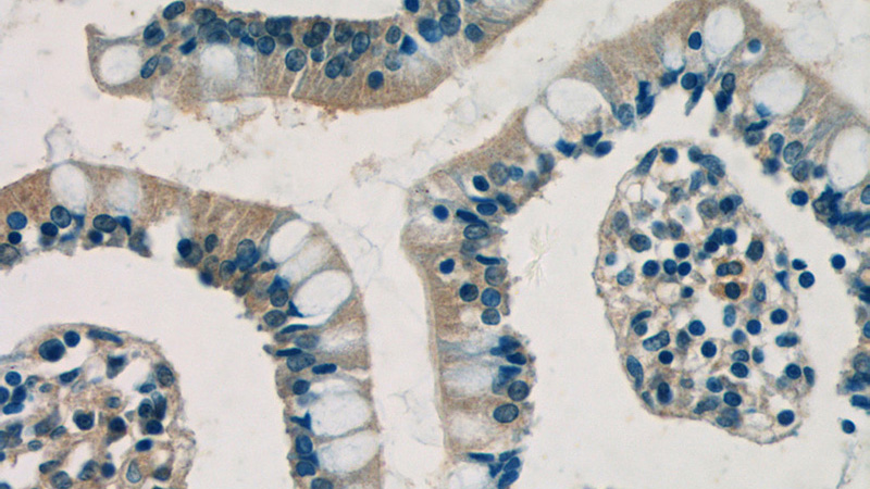
Immunohistochemistry of paraffin-embedded human small intestine tissue slide using Catalog No:115264(SHARPIN Antibody) at dilution of 1:50 (under 40x lens)
-
Background
SHARPIN is as a third component of the linear ubiquitin chain assembly complex (LUBAC), recruited to the CD40 and TNF receptor signaling complexes together with its other constituents, HOIL1 and HOIP(PMID:21455173).SHARPIN is enriched in the postsynaptic density and forms a complex with SHANK in heterologous cells and brain(PMID:11178875).It has 2 isoforms produced by alternative splicing.
-
References
- Rickard JA, Anderton H, Etemadi N. TNFR1-dependent cell death drives inflammation in Sharpin-deficient mice. eLife. 3:. 2014.
- Boisson B, Laplantine E, Dobbs K. Human HOIP and LUBAC deficiency underlies autoinflammation, immunodeficiency, amylopectinosis, and lymphangiectasia. The Journal of experimental medicine. 212(6):939-51. 2015.
- de Almagro MC, Goncharov T, Newton K, Vucic D. Cellular IAP proteins and LUBAC differentially regulate necrosome-associated RIP1 ubiquitination. Cell death & disease. 6:e1800. 2015.
- Peltzer N, Rieser E, Taraborrelli L. HOIP deficiency causes embryonic lethality by aberrant TNFR1-mediated endothelial cell death. Cell reports. 9(1):153-65. 2014.
- Damgaard RB, Nachbur U, Yabal M. The ubiquitin ligase XIAP recruits LUBAC for NOD2 signaling in inflammation and innate immunity. Molecular cell. 46(6):746-58. 2012.
- Douglas T, Champagne C, Morizot A, Lapointe JM, Saleh M. The Inflammatory Caspases-1 and -11 Mediate the Pathogenesis of Dermatitis in Sharpin-Deficient Mice. Journal of immunology (Baltimore, Md. : 1950). 195(5):2365-73. 2015.
- Boisson B, Laplantine E, Prando C. Immunodeficiency, autoinflammation and amylopectinosis in humans with inherited HOIL-1 and LUBAC deficiency. Nature immunology. 13(12):1178-86. 2012.
- Keusekotten K, Elliott PR, Glockner L. OTULIN antagonizes LUBAC signaling by specifically hydrolyzing Met1-linked polyubiquitin. Cell. 153(6):1312-26. 2013.
Related Products / Services
Please note: All products are "FOR RESEARCH USE ONLY AND ARE NOT INTENDED FOR DIAGNOSTIC OR THERAPEUTIC USE"