
-
Product Name
SEPT2 antibody
- Documents
-
Description
SEPT2 Rabbit Polyclonal antibody. Positive WB detected in HeLa cells, C6 cells, human skeletal muscle tissue, Jurkat cells, K-562 cells, mouse brain tissue, mouse heart tissue, mouse testis tissue, rat brain tissue. Positive IP detected in mouse brain tissue. Positive FC detected in MCF-7 cells. Positive IF detected in HepG2 cells, hTERT-RPE1 cells and Mouse embryonic fibroblasts. Positive IHC detected in human pancreas cancer tissue, human liver cancer tissue. Observed molecular weight by Western-blot: 45 kDa
-
Tested applications
ELISA, IHC, IP, IF, WB, FC
-
Species reactivity
Human,Mouse,Rat; other species not tested.
-
Alternative names
DIFF6 antibody; hNedd5 antibody; KIAA0158 antibody; NEDD 5 antibody; NEDD5 antibody; Pnutl3 antibody; SEPT2 antibody; septin 2 antibody; septin2 antibody
-
Isotype
Rabbit IgG
-
Preparation
This antibody was obtained by immunization of SEPT2 recombinant protein (Accession Number: NM_001008492). Purification method: Antigen affinity purified.
-
Clonality
Polyclonal
-
Formulation
PBS with 0.1% sodium azide and 50% glycerol pH 7.3.
-
Storage instructions
Store at -20℃. DO NOT ALIQUOT
-
Applications
Recommended Dilution:
WB: 1:500-1:5000
IP: 1:500-1:5000
IHC: 1:20-1:200
IF: 1:20-1:200
-
Validations
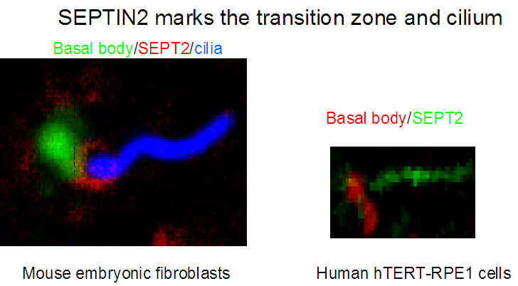
IF result from Dr. Kevin Corbit. SEPT2(Catalog No:115117) marks the transition zone and cilium of Human hTERT-RPE1 cells and Mouse embryonic fibroblasts.
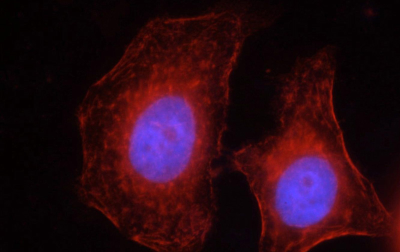
Immunofluorescent analysis of HepG2 cells, using SEPT2 antibody 113971-AP at 1:50 dilution and Rhodamine-labeled goat anti-rabbit IgG (red). Blue pseudocolor = DAPI (fluorescent DNA dye).
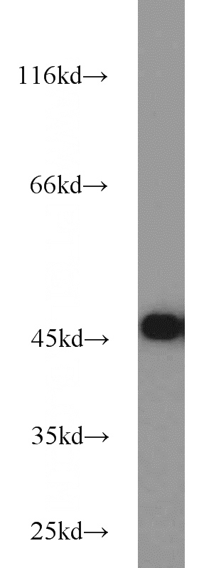
HeLa cells were subjected to SDS PAGE followed by western blot with Catalog No:115117(SEPT2 antibody) at dilution of 1:1000
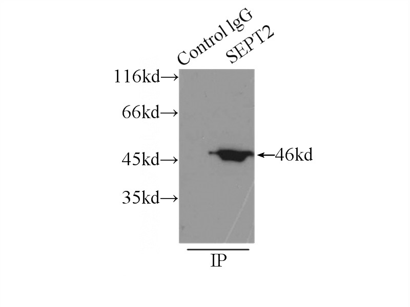
IP Result of anti-SEPT2 (IP:Catalog No:115117, 3ug; Detection:Catalog No:115117 1:1000) with mouse brain tissue lysate 5000ug.
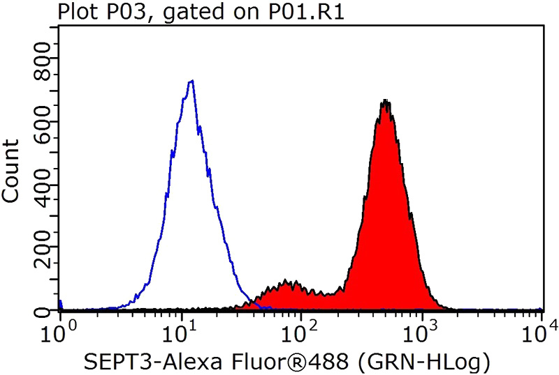
1X10^6 MCF-7 cells were stained with 0.2ug SEPT2 antibody (Catalog No:115117, red) and control antibody (blue). Fixed with 90% MeOH blocked with 3% BSA (30 min). Alexa Fluor 488-congugated AffiniPure Goat Anti-Rabbit IgG(H+L) with dilution 1:1000.
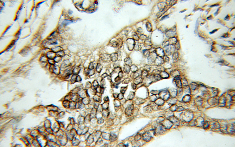
Immunohistochemical of paraffin-embedded human pancreas cancer using Catalog No:115117(SEPT2 antibody) at dilution of 1:50 (under 10x lens)
-
Background
SEPT2, also named as DIFF6, KIAA0158, NEDD5 and Septin-2, belongs to the septin family. It is a filament-forming cytoskeletal GTPase. SEPT2 is required for normal organization of the actin cytoskeleton. It plays a role in ciliogenesis and collective cell movements. SEPT2 is coordinated expression with SEPT6 and SEPT7. (PMID:21737677)
-
References
- Lu Y, Cai G, Cui S. FHL2-driven molecular network mediated Septin2 knockdown inducing apoptosis in mesangial cell. Proteomics. 14(21-22):2485-97. 2014.
- Volceanov L, Herbst K, Biniossek M. Septins arrange F-actin-containing fibers on the Chlamydia trachomatis inclusion and are required for normal release of the inclusion by extrusion. mBio. 5(5):e01802-14. 2014.
- Menon MB, Sawada A, Chaturvedi A. Genetic deletion of SEPT7 reveals a cell type-specific role of septins in microtubule destabilization for the completion of cytokinesis. PLoS genetics. 10(8):e1004558. 2014.
- Kuo YC, Shen YR, Chen HI. SEPT12 orchestrates the formation of mammalian sperm annulus by organizing core octameric complexes with other SEPT proteins. Journal of cell science. 128(5):923-34. 2015.
- Sellin ME, Sandblad L, Stenmark S, Gullberg M. Deciphering the rules governing assembly order of mammalian septin complexes. Molecular biology of the cell. 22(17):3152-64. 2011.
Related Products / Services
Please note: All products are "FOR RESEARCH USE ONLY AND ARE NOT INTENDED FOR DIAGNOSTIC OR THERAPEUTIC USE"