
-
Product Name
RPS3A antibody
- Documents
-
Description
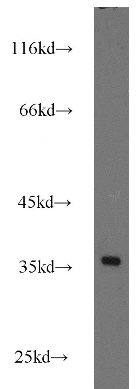
RPS3A Rabbit Polyclonal antibody. Positive IF detected in HepG2 cells. Positive IHC detected in human pancreas cancer tissue. Positive FC detected in HepG2 cells. Positive WB detected in HeLa cells, A431 cells, A549 cells, HepG2 cells, Jurkat cells, Raji cells. Positive IP detected in HepG2 cells. Observed molecular weight by Western-blot: 35kd
-
Tested applications
ELISA, WB, IHC, IF, FC, IP
-
Species reactivity
Human,Mouse,Rat; other species not tested.
-
Alternative names
40S ribosomal protein S3a antibody; Fte 1 antibody; FTE1 antibody; MFTL antibody; ribosomal protein S3A antibody; RPS3A antibody
-
Isotype
Rabbit IgG
-
Preparation
This antibody was obtained by immunization of RPS3A recombinant protein (Accession Number: NM_001006). Purification method: Antigen affinity purified.
-
Clonality
Polyclonal
-
Formulation
PBS with 0.02% sodium azide and 50% glycerol pH 7.3.
-
Storage instructions
Store at -20℃. DO NOT ALIQUOT
-
Applications
Recommended Dilution:
WB: 1:200-1:2000
IP: 1:200-1:2000
IHC: 1:20-1:200
IF: 1:20-1:200
-
Validations
HeLa cells were subjected to SDS PAGE followed by western blot with Catalog No:114907(RPS3A antibody) at dilution of 1:800
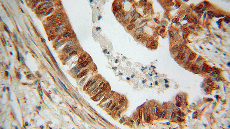
Immunohistochemical of paraffin-embedded human pancreas cancer using Catalog No:114907(RPS3A antibody) at dilution of 1:100 (under 40x lens)
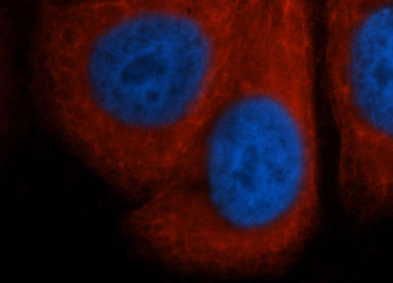
Immunofluorescent analysis of HepG2 cells, using RPS3A antibody Catalog No:114907 at 1:50 dilution and Rhodamine-labeled goat anti-rabbit IgG (red). Blue pseudocolor = DAPI (fluorescent DNA dye).
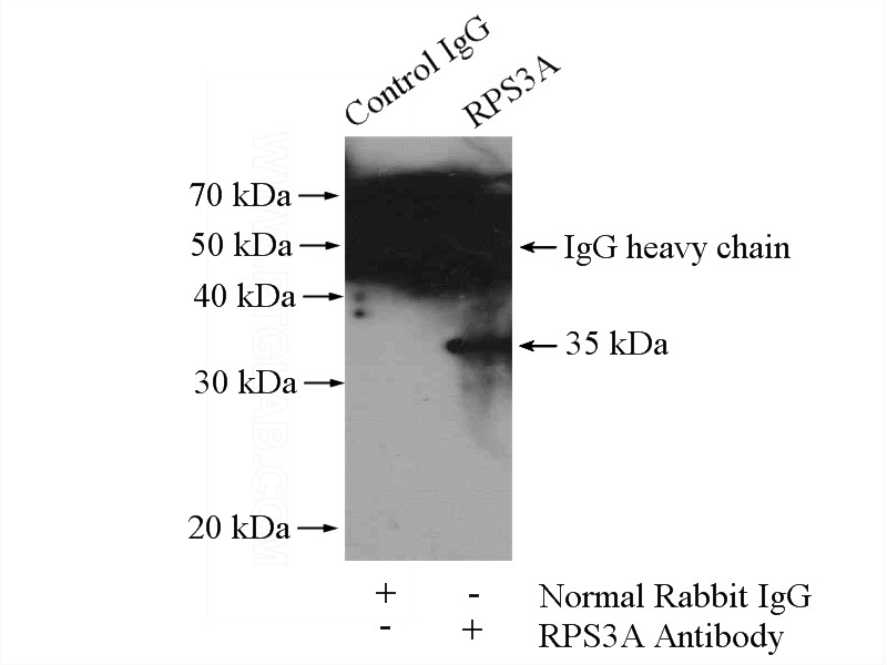
IP Result of anti-RPS3A (IP:Catalog No:114907, 4ug; Detection:Catalog No:114907 1:500) with HepG2 cells lysate 3600ug.
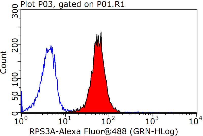
1X10^6 HepG2 cells were stained with 0.2ug RPS3A antibody (Catalog No:114907, red) and control antibody (blue). Fixed with 90% MeOH blocked with 3% BSA (30 min). Alexa Fluor 488-congugated AffiniPure Goat Anti-Rabbit IgG(H+L) with dilution 1:1500.
-
Background
RPS3a, a component of the ribosomal small subunit (40S), is a member of the A20 family of the deubiquitinating cysteine proteases. RPS3a is an inducing factor for oncogenic cellular transformation of Rat-1 cells by v-fos, and is highly expressed in most tumors including hepatocellular carcinoma and other cancers. On the other hand, RPS3a has also been described as a regulator of apoptosis and transcription factors
-
References
- Qi T, Zhang W, Luan Y. Proteomic profiling identified multiple short-lived members of the central proteome as the direct targets of the addicted oncogenes in cancer cells. Molecular & cellular proteomics : MCP. 13(1):49-62. 2014.
Related Products / Services
Please note: All products are "FOR RESEARCH USE ONLY AND ARE NOT INTENDED FOR DIAGNOSTIC OR THERAPEUTIC USE"