
-
Product Name
PDCD6IP antibody
- Documents
-
Description
PDCD6IP Rabbit Polyclonal antibody. Positive IP detected in Jurkat cells. Positive WB detected in Jurkat cells, mouse liver tissue. Positive IF detected in Hela cells. Observed molecular weight by Western-blot: 96kd
-
Tested applications
ELISA, IF, IP, WB
-
Species reactivity
Human,Mouse,Rat; other species not tested.
-
Alternative names
AIP1 antibody; ALG 2 interacting protein 1 antibody; Alix antibody; DRIP4 antibody; HP95 antibody; KIAA1375 antibody; PDCD6 interacting protein antibody; PDCD6IP antibody
-
Isotype
Rabbit IgG
-
Preparation
This antibody was obtained by immunization of PDCD6IP recombinant protein (Accession Number: NM_013374). Purification method: Antigen affinity purified.
-
Clonality
Polyclonal
-
Formulation
PBS with 0.1% sodium azide and 50% glycerol pH 7.3.
-
Storage instructions
Store at -20℃. DO NOT ALIQUOT
-
Applications
Recommended Dilution:
WB: 1:500-1:5000
IP: 1:200-1:2000
IF: 1:10-1:100
-
Validations
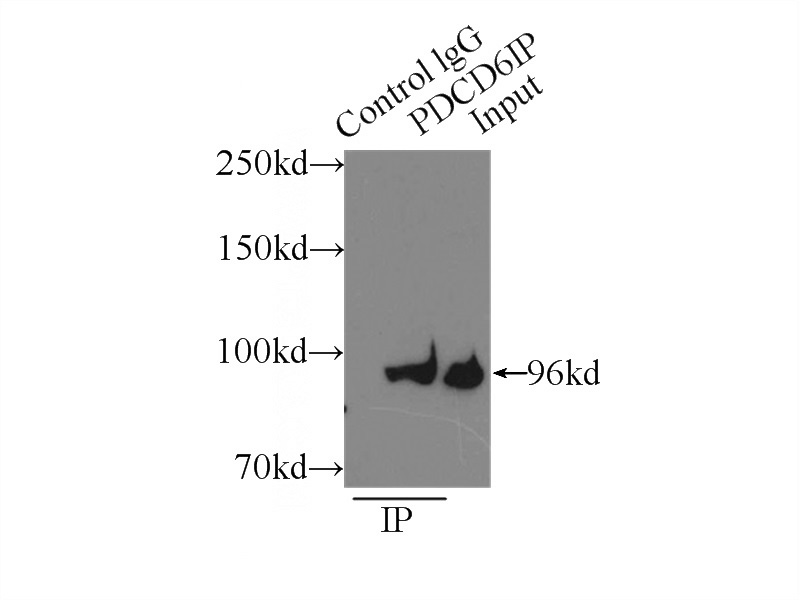
IP Result of anti-ALIX; AIP1 (IP:Catalog No:113768, 3ug; Detection:Catalog No:113768 1:500) with Jurkat cells lysate 4000ug.
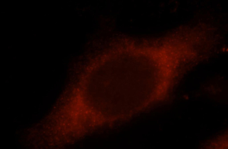
Immunofluorescent analysis of Hela cells, using PDCD6IP antibody Catalog No:113768 at 1:25 dilution and Rhodamine-labeled goat anti-rabbit IgG (red).
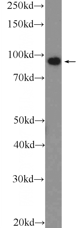
Jurkat cells were subjected to SDS PAGE followed by western blot with Catalog No:113768(ALIX; AIP1 Antibody) at dilution of 1:1000
-
Background
ALG-2-interacting protein 1 (ALIX), also known as AIP1 or Hp95, is encoded by PDCD6IP gene and is involved in cell death through mechanisms involving its binding partner ALG-2 (apoptosis-linked gene-2). ALG-2 is a 22-kDa protein containing five serially repetitive EF-hand structures and is defined as a regulator of calcium-induced apoptosis following endoplasmic reticulum (ER) stress. ALIX interacts with ALG-2 through its C-terminal proline-rich region and participates in formation of multivesicular bodies. Recent finding suggest that ALIX is a critical component of caspase 9 activation and apoptosis triggered by calcium.
-
References
- Hopp K, Ward CJ, Hommerding CJ. Functional polycystin-1 dosage governs autosomal dominant polycystic kidney disease severity. The Journal of clinical investigation. 122(11):4257-73. 2012.
- Liang B, Peng P, Chen S. Characterization and proteomic analysis of ovarian cancer-derived exosomes. Journal of proteomics. 80:171-82. 2013.
- Li J, Liu K, Liu Y. Exosomes mediate the cell-to-cell transmission of IFN-α-induced antiviral activity. Nature immunology. 14(8):793-803. 2013.
- Zhang W, Peng P, Kuang Y. Characterization of exosomes derived from ovarian cancer cells and normal ovarian epithelial cells by nanoparticle tracking analysis. Tumour biology : the journal of the International Society for Oncodevelopmental Biology and Medicine. 2015.
Related Products / Services
Please note: All products are "FOR RESEARCH USE ONLY AND ARE NOT INTENDED FOR DIAGNOSTIC OR THERAPEUTIC USE"