

-
Product Name
OAS2 antibody
- Documents
-
Description
OAS2 Rabbit Polyclonal antibody. Positive IHC detected in human breast cancer tissue, human colon tissue, human liver cancer tissue. Positive IF detected in HepG2 cells. Positive WB detected in Jurkat cells, HL-60 cells, human liver tissue, mouse lung tissue. Positive IP detected in Jurkat cells. Observed molecular weight by Western-blot: 66-71 kDa
-
Tested applications
ELISA, WB, IHC, IF, IP
-
Species reactivity
Human, Mouse; other species not tested.
-
Alternative names
(2 5)oligo(A) synthase 2 antibody; 2 5 oligoadenylate synthase 2 antibody; 2 5A synthase 2 antibody; OAS2 antibody; p69 OAS / p71 OAS antibody; p69OAS / p71OAS antibody
-
Isotype
Rabbit IgG
-
Preparation
This antibody was obtained by immunization of OAS2 recombinant protein (Accession Number: NM_002535). Purification method: Antigen affinity purified.
-
Clonality
Polyclonal
-
Formulation
PBS with 0.02% sodium azide and 50% glycerol pH 7.3.
-
Storage instructions
Store at -20℃. DO NOT ALIQUOT
-
Applications
Recommended Dilution:
WB: 1:500-1:5000
IP: 1:200-1:2000
IHC: 1:20-1:200
IF: 1:10-1:100
-
Validations
Jurkat cells were subjected to SDS PAGE followed by western blot with Catalog No:113460(OAS2 antibody) at dilution of 1:500
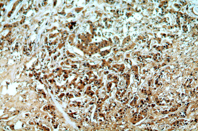
Immunohistochemical of paraffin-embedded human breast cancer using Catalog No:113460(OAS2 antibody) at dilution of 1:50 (under 10x lens)
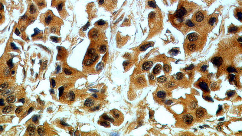
Immunohistochemical of paraffin-embedded human breast cancer using Catalog No:113460(OAS2 antibody) at dilution of 1:50 (under 40x lens)
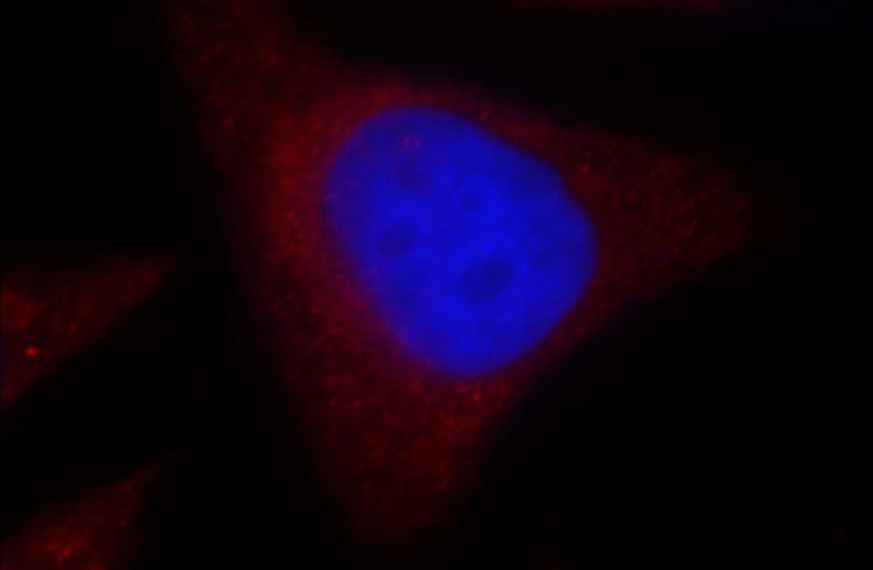
Immunofluorescent analysis of HepG2 cells, using OAS2 antibody Catalog No:113460 at 1:25 dilution and Rhodamine-labeled goat anti-rabbit IgG (red). Blue pseudocolor = DAPI (fluorescent DNA dye).
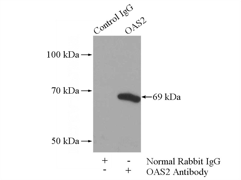
IP Result of anti-OAS2 (IP:Catalog No:113460, 4ug; Detection:Catalog No:113460 1:500) with Jurkat cells lysate 3600ug.
-
Background
The 2-prime,5-prime oligoadenylate synthetases (OASs), such as OAS2, are interferon-induced proteins characterized by their capacity to catalyze the synthesis of 2-prime,5-prime oligomers of adenosine (2-5As). OAS2 is also named as p69 OAS / p71 OAS. It has 3 isoforms(71/69/20 kDa) produced by alternative splicing. The OAS2 isozymes are 69–71 kDa and form dimers(PMID:17765707). Glycosylation is essential for its activity(PMID:9880569). Constitutive expression of the 69-kDa form of the OAS2 protein in human cells inhibited the replication of encephalomyocarditis virus (EMCV) but not that of VSV, Sendai virus, or reovirus(PMID:16501108). This antibody is specific to isoform p71 and isoform p69 of OAS2.
-
References
- Grammatikos AP, Ghosh D, Devlin A, Kyttaris VC, Tsokos GC. Spleen tyrosine kinase (Syk) regulates systemic lupus erythematosus (SLE) T cell signaling. PloS one. 8(8):e74550. 2013.
Related Products / Services
Please note: All products are "FOR RESEARCH USE ONLY AND ARE NOT INTENDED FOR DIAGNOSTIC OR THERAPEUTIC USE"