
-
Product Name
N-cadherin antibody
- Documents
-
Description
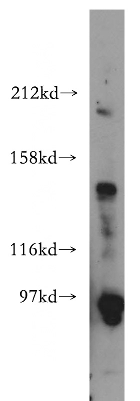
N-cadherin Rabbit Polyclonal antibody. Positive IHC detected in human heart tissue, mouse brain tissue. Positive WB detected in HEK-293 cells, mouse heart tissue. Observed molecular weight by Western-blot: 90kd,140kd
-
Tested applications
ELISA, WB, IHC
-
Species reactivity
Human,Mouse,Rat; other species not tested.
-
Alternative names
Cadherin 2 antibody; CD325 antibody; CDH2 antibody; CDHN antibody; CDw325 antibody; N cadherin antibody; NCAD antibody; N-cadherin antibody; Neural cadherin antibody
-
Isotype
Rabbit IgG
-
Preparation
This antibody was obtained by immunization of N-cadherin recombinant protein (Accession Number: NM_001792). Purification method: Antigen affinity purified.
-
Clonality
Polyclonal
-
Formulation
PBS with 0.02% sodium azide and 50% glycerol pH 7.3.
-
Storage instructions
Store at -20℃. DO NOT ALIQUOT
-
Applications
Recommended Dilution:
WB: 1:200-1:2000
IHC: 1:20-1:200
-
Validations
HEK-293 cells were subjected to SDS PAGE followed by western blot with Catalog No:113025(N-cadherin antibody) at dilution of 1:500
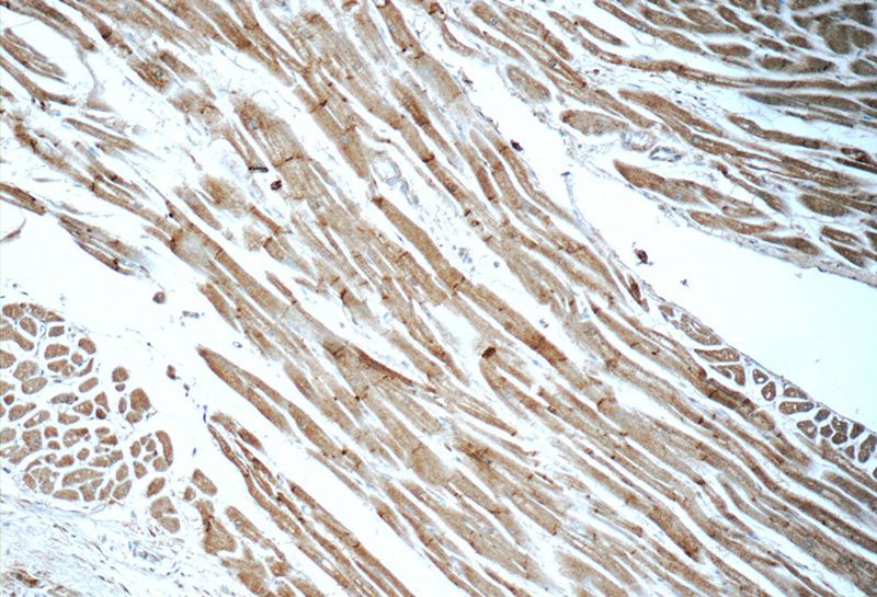
Immunohistochemistry of paraffin-embedded human heart tissue slide using Catalog No:113025(N-cadherin Antibody) at dilution of 1:50 (under 10x lens)
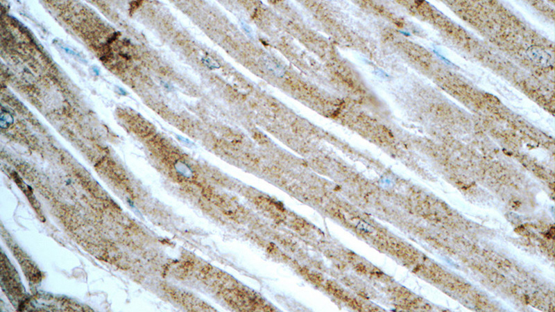
Immunohistochemistry of paraffin-embedded human heart tissue slide using Catalog No:113025(N-cadherin Antibody) at dilution of 1:50 (under 40x lens)
-
Background
Cadherins are a family of transmembrane glycoproteins that mediate calcium-dependent cell-cell adhesion and play an important role in the maintenance of normal tissue architecture. N-cadherin (neural cadherin), also known as CDH2 (cadherin 2), is a classical member of the cadherin superfamily which also include E-, P-, R-, and B-cadherins. Expression of N-cadherin has been reported on various cell types including neurons, endothelial cells and cardiac myocytes (PMID: 11282032; 9508779; 8125202). N-cadherin has functions in early brain morphogenesis, synaptogenesis and synaptic plasticity (PMID: 23321619).
-
References
- Ning Q, Liu C, Hou L. Vascular endothelial growth factor receptor-1 activation promotes migration and invasion of breast cancer cells through epithelial-mesenchymal transition. PloS one. 8(6):e65217. 2013.
- Liu N, Li Y, Su S, Wang N, Wang H, Li J. Inhibition of cell migration by ouabain in the A549 human lung cancer cell line. Oncology letters. 6(2):475-479. 2013.
- Teng Y, Zhao L, Zhang Y, Chen W, Li X. Id-1, a protein repressed by miR-29b, facilitates the TGFβ1-induced epithelial-mesenchymal transition in human ovarian cancer cells. Cellular physiology and biochemistry : international journal of experimental cellular physiology, biochemistry, and pharmacology. 33(3):717-30. 2014.
- Li XL, Dong X, Xue Y, Li CF, Gou WL, Chen Q. Increased expression levels of E-cadherin, cytokeratin 18 and 19 observed in preeclampsia were not correlated with disease severity. Placenta. 35(8):625-31. 2014.
- McLane JS, Rivet CJ, Gilbert RJ, Ligon LA. A biomaterial model of tumor stromal microenvironment promotes mesenchymal morphology but not epithelial to mesenchymal transition in epithelial cells. Acta biomaterialia. 10(11):4811-21. 2014.
- Hu TH, Yao Y, Yu S. SDF-1/CXCR4 promotes epithelial-mesenchymal transition and progression of colorectal cancer by activation of the Wnt/β-catenin signaling pathway. Cancer letters. 354(2):417-26. 2014.
- Huang W, Liu J, Feng X. DLC-1 induces mitochondrial apoptosis and epithelial mesenchymal transition arrest in nasopharyngeal carcinoma by targeting EGFR/Akt/NF-κB pathway. Medical oncology (Northwood, London, England). 32(4):115. 2015.
- Song L, Liu D, Wang B. miR-494 suppresses the progression of breast cancer in vitro by targeting CXCR4 through the Wnt/β-catenin signaling pathway. Oncology reports. 34(1):525-31. 2015.
Related Products / Services
Please note: All products are "FOR RESEARCH USE ONLY AND ARE NOT INTENDED FOR DIAGNOSTIC OR THERAPEUTIC USE"