
-
Product Name
MYBBP1A antibody
- Documents
-
Description
MYBBP1A Rabbit Polyclonal antibody. Positive FC detected in HepG2 cells. Positive IHC detected in human kidney tissue. Positive IF detected in HepG2 cells. Positive WB detected in HeLa cells, HEK-293 cells. Positive IP detected in HEK-293 cells. Observed molecular weight by Western-blot: 160kd
-
Tested applications
ELISA, WB, IHC, IF, FC, IP
-
Species reactivity
Human; other species not tested.
-
Alternative names
FLJ37886 antibody; MYB binding protein (P160) 1a antibody; Myb binding protein 1A antibody; MYBBP1A antibody; P160 antibody; PAP2 antibody
-
Isotype
Rabbit IgG
-
Preparation
This antibody was obtained by immunization of MYBBP1A recombinant protein (Accession Number: NM_014520). Purification method: Antigen affinity purified.
-
Clonality
Polyclonal
-
Formulation
PBS with 0.02% sodium azide and 50% glycerol pH 7.3.
-
Storage instructions
Store at -20℃. DO NOT ALIQUOT
-
Applications
Recommended Dilution:
WB: 1:500-1:5000
IP: 1:200-1:2000
IHC: 1:20-1:200
IF: 1:20-1:200
-
Validations
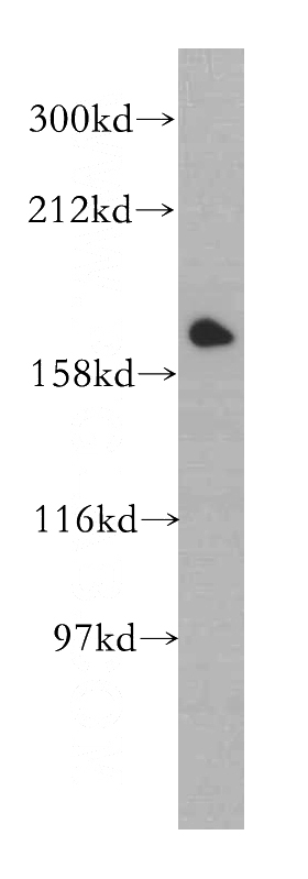
HeLa cells were subjected to SDS PAGE followed by western blot with Catalog No:112912(MYBBP1A antibody) at dilution of 1:500
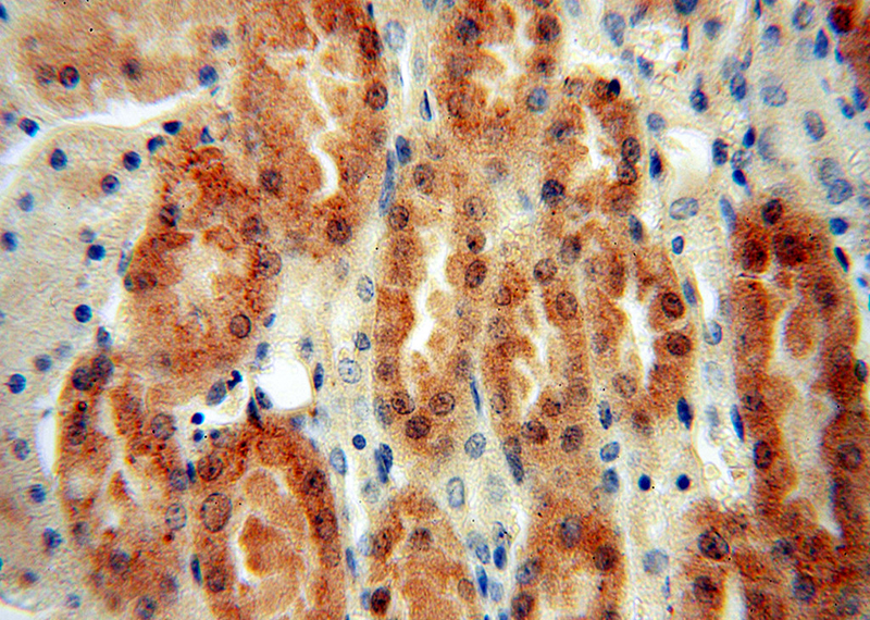
Immunohistochemical of paraffin-embedded human kidney using Catalog No:112912(MYBBP1A antibody) at dilution of 1:100 (under 40x lens)
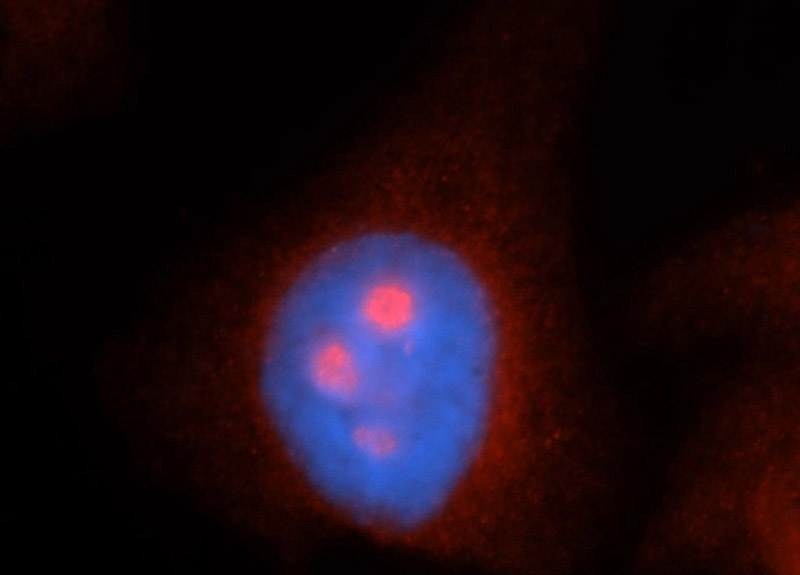
Immunofluorescent analysis of HepG2 cells, using MYBBP1A antibody Catalog No:112912 at 1:50 dilution and Rhodamine-labeled goat anti-rabbit IgG (red). Blue pseudocolor = DAPI (fluorescent DNA dye).
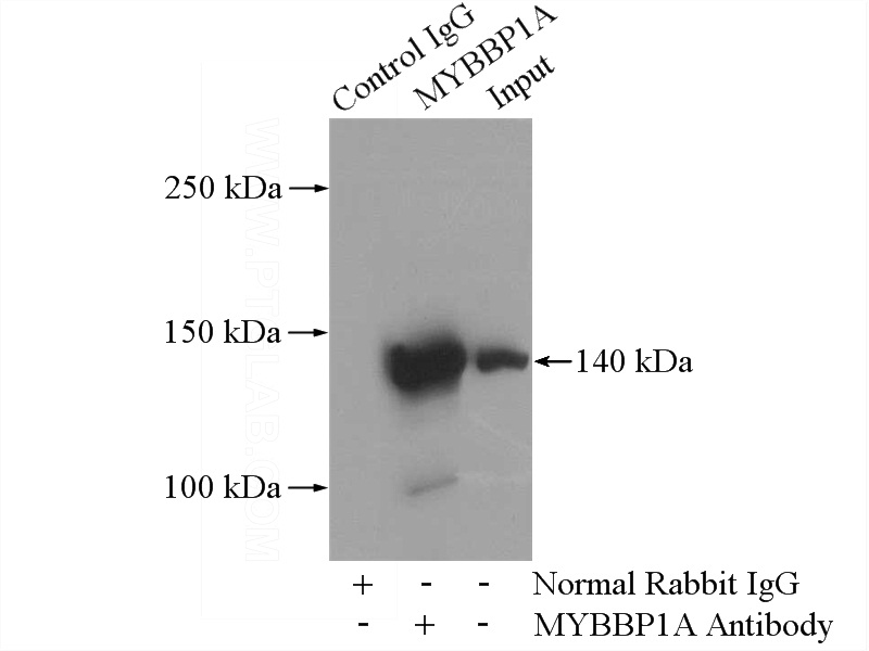
IP Result of anti-MYBBP1A (IP:Catalog No:112912, 4ug; Detection:Catalog No:112912 1:500) with HEK-293 cells lysate 2800ug.
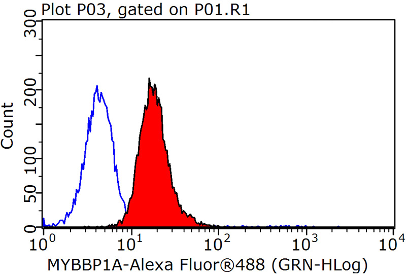
1X10^6 HepG2 cells were stained with 0.2ug MYBBP1A antibody (Catalog No:112912, red) and control antibody (blue). Fixed with 90% MeOH blocked with 3% BSA (30 min). Alexa Fluor 488-congugated AffiniPure Goat Anti-Rabbit IgG(H+L) with dilution 1:1500.
-
Background
The protooncogene MYB is predominantly expressed in immature hemopoietic cells where it has an essential role in hemopoietic cell proliferation and differentiation. Oncogenically activated forms of MYB is generally N- and/or C-terminal truncations of the normal MYB protein. Removal of the C terminus of MYB disrupts or deletes a region termed the negative regulatory domain (NRD), resulting in an increase in DNA binding, transactivation, and transformation by MYB. One feature of the NRD is a leucine zipper-like motif [PMID: 8302594]. Murine Myb-binding protein-1a (MYBBP1A), originally called P160, was identified by its ability to interact specifically with Myb via this leucine zipper-like motif. MYBBP1A modulates MYB activity upon binding to the MYB NRD [PMID: 10644447, 9447996].
Related Products / Services
Please note: All products are "FOR RESEARCH USE ONLY AND ARE NOT INTENDED FOR DIAGNOSTIC OR THERAPEUTIC USE"