
-
Product Name
MTUS1 antibody
- Documents
-
Description
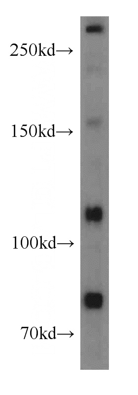
MTUS1 Rabbit Polyclonal antibody. Positive IF detected in PC-3 cells. Positive IP detected in mouse brain tissue. Positive WB detected in Jurkat cells, human brain tissue, K-562 cells, PC-3 cells. Observed molecular weight by Western-blot: 40 kDa,80 kDa,110-120 kDa,270-280 kDa
-
Tested applications
ELISA, WB, IP, IF
-
Species reactivity
Human,Mouse,Rat; other species not tested.
-
Alternative names
AT2 receptor binding protein antibody; ATBP antibody; ATIP antibody; DKFZp586D1519 antibody; DKFZp686F20243 antibody; FLJ14295 antibody; GK1 antibody; KIAA1288 antibody; MP44 antibody; MTSG1 antibody; MTUS1 antibody
-
Isotype
Rabbit IgG
-
Preparation
This antibody was obtained by immunization of MTUS1 recombinant protein (Accession Number: BC033842). Purification method: Antigen affinity purified.
-
Clonality
Polyclonal
-
Formulation
PBS with 0.02% sodium azide and 50% glycerol pH 7.3.
-
Storage instructions
Store at -20℃. DO NOT ALIQUOT
-
Applications
Recommended Dilution:
WB: 1:500-1:5000
IP: 1:200-1:2000
IF: 1:20-1:200
-
Validations
Jurkat cells were subjected to SDS PAGE followed by western blot with Catalog No:112883(MTUS1 antibody) at dilution of 1:500
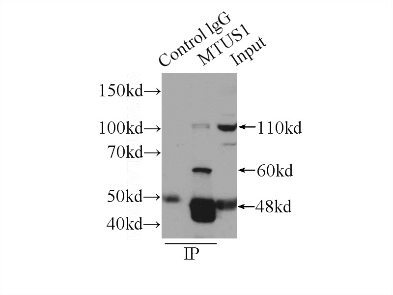
IP Result of anti-MTUS1 (IP:Catalog No:112883, 4ug; Detection:Catalog No:112883 1:500) with mouse brain tissue lysate 6000ug.
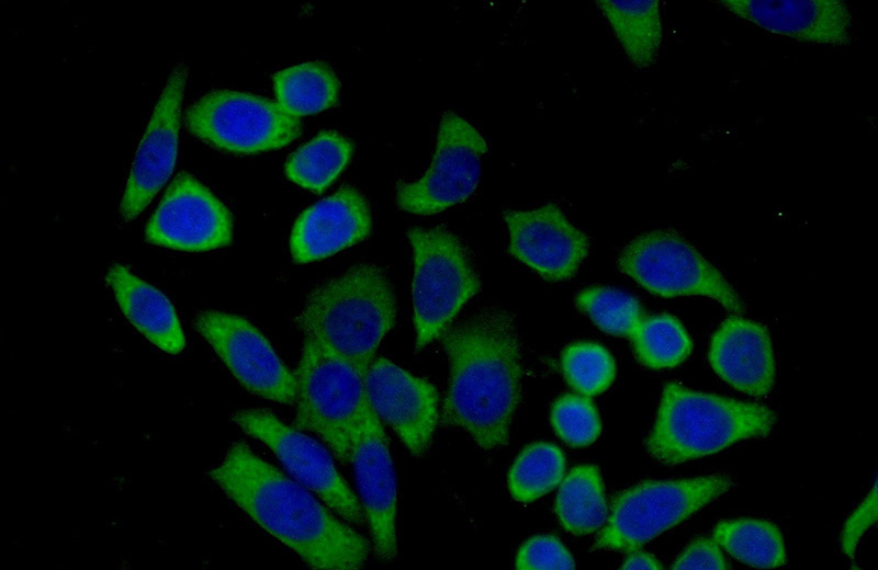
Immunofluorescent analysis of (-20oc Ethanol) fixed PC-3 cells using Catalog No:112883(MTUS1 Antibody) at dilution of 1:50 and Alexa Fluor 488-congugated AffiniPure Goat Anti-Rabbit IgG(H+L)
-
Background
MTUS1, also named as ATBP, ATIP, GK1, KIAA1288 and MTSG1, Belongs to the MTUS1 family. It cooperates with AGTR2 to inhibit ERK2 activation and cell proliferation. MTUS1 may be required for AGTR2 cell surface expression. Together with PTPN6, induces UBE2V2 expression upon angiotensin-II stimulation. Isoform 1 inhibits breast cancer cell proliferation, delays the progression of mitosis by prolonging metaphase and reduces tumor growth. MTUS1 up-regulation during cellular transition from proliferation to quiescence and differentiation. It is a potential tumor suppressor gene located at chromosome 8p21.3.22, near marker D8S254. According to the functional data and intracellular localization, MTUS1 also named as mitochondrial tumor suppressor gene 1 (MTSG1). One main feature common to all ATIP members is the presence of a large C-terminal coiled-coil domain that allows homo- and hetero-dimerization of these proteins. (PMID:12692079, 15123706) The antibody can recognize all the isoforms except isoform 5 (85-90kd). The antibody tested HomoDimer isoforms (80kd/110-120/280kd) in Jurkat cell.
Related Products / Services
Please note: All products are "FOR RESEARCH USE ONLY AND ARE NOT INTENDED FOR DIAGNOSTIC OR THERAPEUTIC USE"