
-
Product Name
MITF antibody
- Documents
-
Description
MITF Rabbit Polyclonal antibody. Positive IP detected in mouse heart tissue. Positive WB detected in Jurkat cells, HeLa cells, mouse heart tissue, rat skin tissue. Positive FC detected in hESC cells. Observed molecular weight by Western-blot: 60-65kd
-
Tested applications
ELISA, WB, FC, IP
-
Species reactivity
Human,Mouse,Rat; other species not tested.
-
Alternative names
bHLHe32 antibody; MITF antibody; WS2A antibody
-
Isotype
Rabbit IgG
-
Preparation
This antibody was obtained by immunization of MITF recombinant protein (Accession Number: BC012503). Purification method: Antigen affinity purified.
-
Clonality
Polyclonal
-
Formulation
PBS with 0.02% sodium azide and 50% glycerol pH 7.3.
-
Storage instructions
Store at -20℃. DO NOT ALIQUOT
-
Applications
Recommended Dilution:
WB: 1:1000-1:10000
IP: 1:500-1:5000
-
Validations
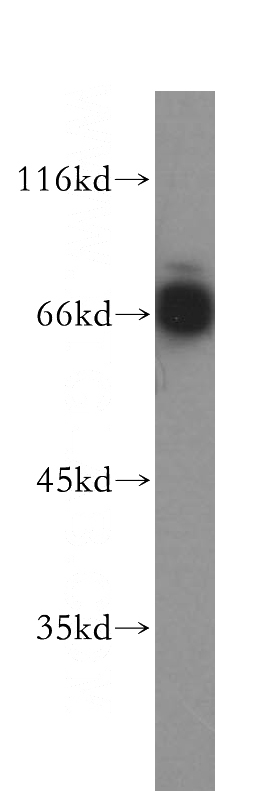
Jurkat cells were subjected to SDS PAGE followed by western blot with Catalog No:112661(MITF antibody) at dilution of 1:400
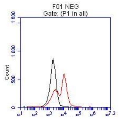
FC result of anti-MITF (Catalog No:112661, 1:50) with hESC which spontaneous differentiation toward retinal lineage (RPE). (Black: control; Red MITF).
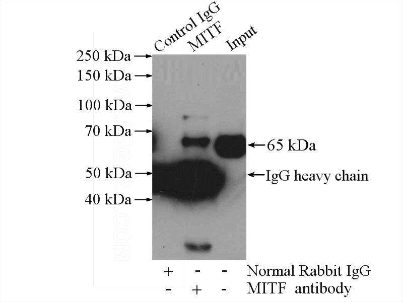
IP Result of anti-MITF (IP:Catalog No:112661, 4ug; Detection:Catalog No:112661 1:1000) with mouse heart tissue lysate 4000ug.
-
Background
The retinal pigment epithelium (RPE) has a essential role in maintaining visual function and dedifferentiation of RPE contributes to the pathophysiology of several ocular diseases[PMID: 22523078]. Microphthalmia-associated transcription factor (MITF) is a key regulator of RPE differentiation that is also down-regulated in dedifferentiated hfRPE cells. MITF is a basic helix-loop-helix (hHLH)-leucine zipper protein that involves in the development of various cell types, including neural crest-derived melanocytes and optic cup-derived retinal pigment epithelial cells [PMID: 10578055].
-
References
- Lee CS, Park M, Han J. Liver X receptor activation inhibits melanogenesis through the acceleration of ERK-mediated MITF degradation. The Journal of investigative dermatology. 133(4):1063-71. 2013.
- Won YM, Seong ZK, Kim JL. Triterpene glycosides with stimulatory activity on melanogenesis from the aerial parts of Weigela subsessilis. Archives of pharmacal research. 38(8):1541-51. 2015.
- Park J, Chung H, Bang SH. (E)-4-(3,4-Dimethoxyphenyl)but-3-en-1-ol Enhances Melanogenesis through Increasing Upstream Stimulating Factor-1-Mediated Tyrosinase Expression. PloS one. 10(11):e0141988. 2015.
Related Products / Services
Please note: All products are "FOR RESEARCH USE ONLY AND ARE NOT INTENDED FOR DIAGNOSTIC OR THERAPEUTIC USE"