
-
Product Name
MCT4 antibody
- Documents
-
Description
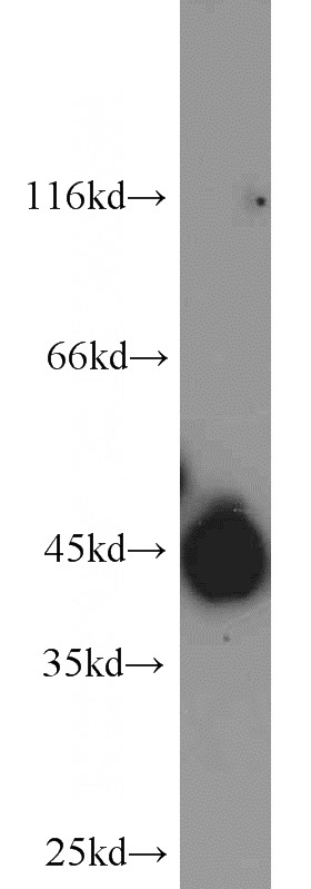
MCT4 Rabbit Polyclonal antibody. Positive WB detected in HeLa cells. Positive FC detected in HepG2 cells. Positive IF detected in HepG2 cells. Positive IHC detected in human breast cancer tissue, human prostate cancer tissue, human skeletal muscle tissue. Observed molecular weight by Western-blot: 42-45 kDa
-
Tested applications
ELISA, WB, IHC, IF, FC
-
Species reactivity
Human; other species not tested.
-
Alternative names
MCT 3 antibody; MCT 4 antibody; MCT3 antibody; MCT4 antibody; MonOCarboxylate transporter 3 antibody; MonOCarboxylate transporter 4 antibody; SLC16A3 antibody
-
Isotype
Rabbit IgG
-
Preparation
This antibody was obtained by immunization of MCT4 recombinant protein (Accession Number: NM_001206951). Purification method: Antigen affinity purified.
-
Clonality
Polyclonal
-
Formulation
PBS with 0.02% sodium azide and 50% glycerol pH 7.3.
-
Storage instructions
Store at -20℃. DO NOT ALIQUOT
-
Applications
Recommended Dilution:
WB: 1:500-1:5000
IHC: 1:20-1:200
IF: 1:20-1:200
-
Validations
HeLa cells were subjected to SDS PAGE followed by western blot with Catalog No:112563(SLC16A3 antibody) at dilution of 1:1000
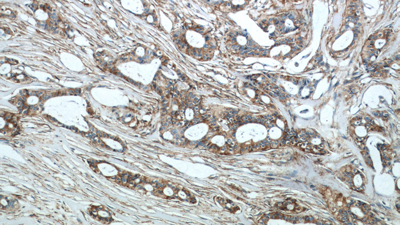
Immunohistochemical of paraffin-embedded human breast cancer using Catalog No:112563(SLC16A3 antibody) at dilution of 1:50 (under 10x lens)
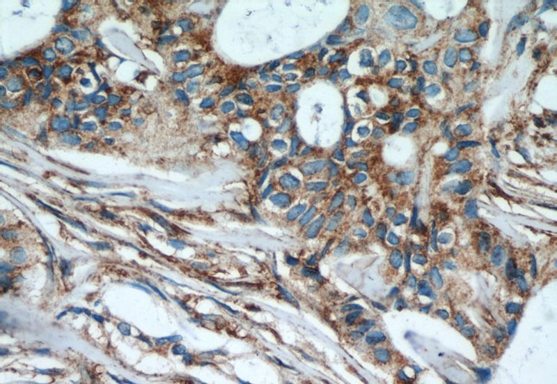
Immunohistochemical of paraffin-embedded human breast cancer using Catalog No:112563(SLC16A3 antibody) at dilution of 1:50 (under 40x lens)
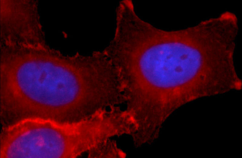
Immunofluorescent analysis of HepG2 cells using Catalog No:112563 (SLC16A3 Antibody) at dilution of 1:50 and Rhodamine-labeled goat anti-rabbit IgG (red).
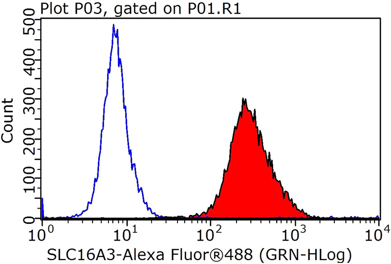
1X10^6 HepG2 cells were stained with 0.2ug SLC16A3 antibody (Catalog No:112563, red) and control antibody (blue). Fixed with 90% MeOH blocked with 3% BSA (30 min). Alexa Fluor 488-congugated AffiniPure Goat Anti-Rabbit IgG(H+L) with dilution 1:1000.
-
Background
The monocarboxylate transporter 4 (MCT4, also known as SLC16A3) is involved in the transportation of metabolically important monocarboxylates such as lactate, pyruvate, acetate and ketone bodies. It is widely expressed, particularly strongly in glycolytic tissues such as white skeletal muscle fibres, astrocytes, white blood cells, chondrocytes and some mammalian cell lines. MCT4 is also linked to tumor biology because it mediates lactate transport across membranes resulting in antiapoptotic effects.
-
References
- Shi H, Jiang H, Wang L. Overexpression of monocarboxylate anion transporter 1 and 4 in T24-induced cancer-associated fibroblasts regulates the progression of bladder cancer cells in a 3D microfluidic device. Cell cycle (Georgetown, Tex.). 14(19):3058-65. 2015.
Related Products / Services
Please note: All products are "FOR RESEARCH USE ONLY AND ARE NOT INTENDED FOR DIAGNOSTIC OR THERAPEUTIC USE"