
-
Product Name
MAP1B antibody
- Documents
-
Description
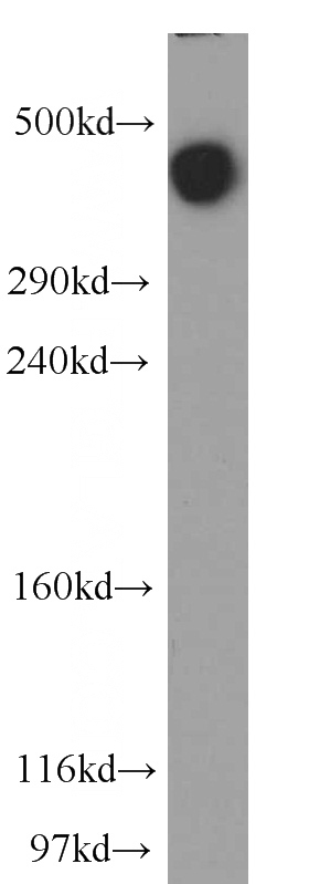
MAP1B Rabbit Polyclonal antibody. Positive WB detected in mouse cerebellum tissue, human brain tissue. Positive FC detected in SH-SY5Y cells. Positive IHC detected in mouse brain tissue. Positive IF detected in SH-SY5Y cells. Observed molecular weight by Western-blot: 320kd
-
Tested applications
ELISA, WB, IHC, FC, IF
-
Species reactivity
Human, Mouse; other species not tested.
-
Alternative names
MAP 1B antibody; MAP1B antibody; MAP5 antibody
- Immunogen
-
Isotype
Rabbit IgG
-
Preparation
This antibody was obtained by immunization of MAP1B recombinant protein (Accession Number: BC141853). Purification method: Antigen affinity purified.
-
Clonality
Polyclonal
-
Formulation
PBS with 0.1% sodium azide and 50% glycerol pH 7.3.
-
Storage instructions
Store at -20℃. DO NOT ALIQUOT
-
Applications
Recommended Dilution:
WB: 1:200-1:2000
IHC: 1:20-1:200
IF: 1:10-1:100
-
Validations
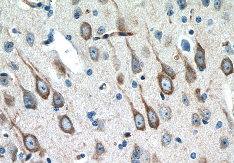
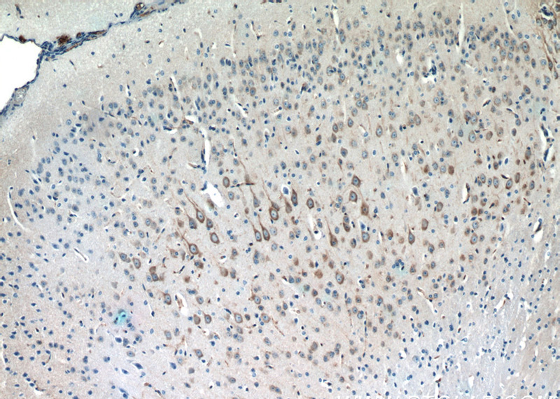
Immunohistochemistry of paraffin-embedded mouse brain tissue slide using Catalog No:112472(MAP1B Antibody) at dilution of 1:50 (under 40x lens)
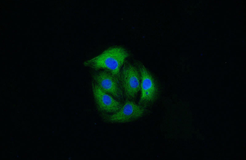
Immunofluorescent analysis of SH-SY5Y cells using Catalog No:112472(MAP1B Antibody) at dilution of 1:25 and Alexa Fluor 488-congugated AffiniPure Goat Anti-Rabbit IgG(H+L)
Immunohistochemistry of paraffin-embedded mouse brain tissue slide using Catalog No:112472(MAP1B Antibody) at dilution of 1:50 (under 10x lens)
mouse cerebellum tissue were subjected to SDS PAGE followed by western blot with Catalog No:112472(MAP1B antibody) at dilution of 1:500
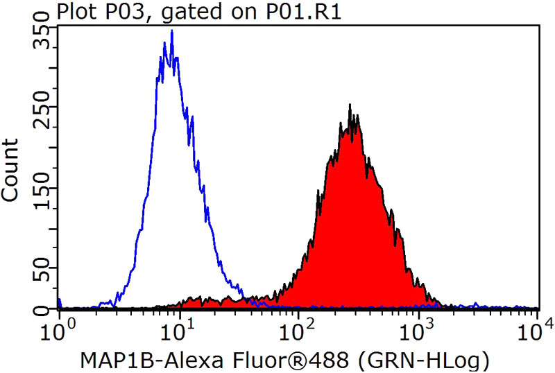
1X10^6 SH-SY5Y cells were stained with 0.2ug MAP1B antibody (Catalog No:112472, red) and control antibody (blue). Fixed with 90% MeOH blocked with 3% BSA (30 min). Alexa Fluor 488-congugated AffiniPure Goat Anti-Rabbit IgG(H+L) with dilution 1:1000.
-
Background
Microtubule-associated protein 1B (MAP1B) is a cytoskeleton protein which can promote microtubule assembly. Previous reports have suggested that this protein is closely involved in neuronal development based on its extensive expression in the developing brain and moderate in mature neurons. Gene disruption or knockout studies of the MAP1B gene led to a delayed development of the nervous system in mice. It includes the N-terminal heavy chain and a C-terminal light chain. The MAP1B heavy chain has a microtubule-stabilization effect, and contains an actin-binding site that may play a role in the crosslinking of actin and microtubules, a function that may be important in neurite elongation. Various isoforms around 300-350 kDa of MAP1B can be observed due to the differences in phosphorylation state. (10704485)
Related Products / Services
Please note: All products are "FOR RESEARCH USE ONLY AND ARE NOT INTENDED FOR DIAGNOSTIC OR THERAPEUTIC USE"