
-
Product Name
LC3B-Specific antibody
- Documents
-
Description
LC3B-Specific Rabbit Polyclonal antibody. Positive FC detected in HeLa cells. Positive IHC detected in human heart tissue, human testis tissue. Positive IF detected in Chloroquine treated HepG2 cells, Starvation treated HEK-293 cells. Positive WB detected in Chloroquine treated HEK-293 cells, HeLa cells, HepG2 cells, human brain tissue, MCF7 cells, mouse brain tissue, rat brain tissue, TN treated Hela cells, UV treated HEK-293 cells. Observed molecular weight by Western-blot: 15kDa, 18 kDa
-
Tested applications
ELISA, IF, IHC, FC, WB
-
Species reactivity
Human, Mouse, Rat; other species not tested.
-
Alternative names
LC3 antibody; LC3B antibody; MAP1A/1BLC3 antibody; MAP1A/MAP1B LC3 B antibody; MAP1A/MAP1B light chain 3 B antibody; MAP1ALC3 antibody; MAP1LC3B antibody
-
Isotype
Rabbit IgG
-
Preparation
This antibody was obtained by immunization of Peptide (Accession Number: NM_022818). Purification method: Antigen affinity purified.
-
Clonality
Polyclonal
-
Formulation
PBS with 0.02% sodium azide and 50% glycerol pH 7.3.
-
Storage instructions
Store at -20℃. DO NOT ALIQUOT
-
Applications
Recommended Dilution:
WB: 1:500-1:5000
IHC: 1:20-1:200
IF: 1:20-1:200
-
Validations
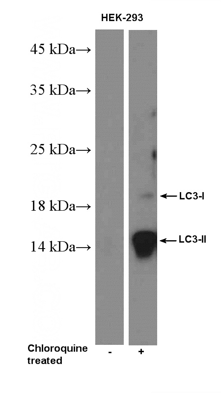
Chloroquine treated HEK-293 cells were subjected to SDS PAGE followed by western blot with Catalog No:112166(MAP1LC3B-Specific Antibody) at dilution of 1:1000
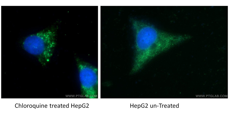
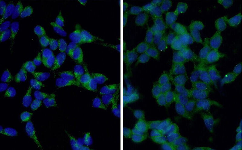
Immunofluorescent analysis of Chloroquine treated/untreated HepG2 cells using Catalog No:112166 (LC3B-Specific Antibody) at dilution of 1:50 and Alexa Fluor 488-congugated AffiniPure Goat Anti-Rabbit IgG(H+L). Cells were fixed with ethanol at -20 oc.
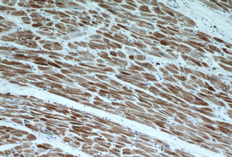
Immunohistochemical of paraffin-embedded human heart using Catalog No:112166 (LC3B-Specific antibody) at dilution of 1:100 (under 10x lens).
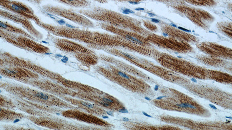
Immunohistochemical of paraffin-embedded human heart using Catalog No:112166 (LC3B-Specific antibody) at dilution of 1:100 (under 40x lens).
Immunofluorescent analysis of starvation treated HEK-293 cells using Catalog No:112166 (LC3B-Specific Antibody) at dilution of 1:50 and Alexa Fluor 488-congugated AffiniPure Goat Anti-Rabbit IgG(H+L). Cells were fixed with ethanol at -20 °C. 20 mM chloroquine was used to block the autophagy flux.
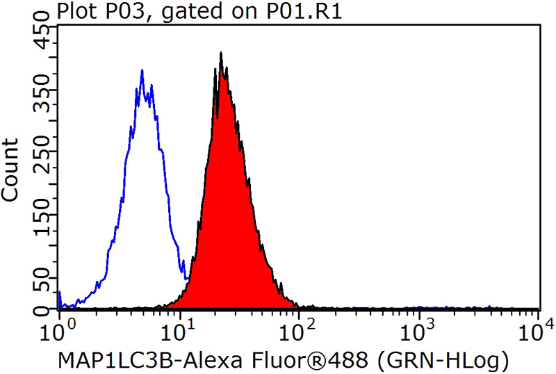
1X10^6 HeLa cells were stained with 0.2ug LC3B-Specific antibody (Catalog No:112166, red) and control antibody (blue). Fixed with 90% MeOH blocked with 3% BSA (30 min). Alexa Fluor 488-congugated AffiniPure Goat Anti-Rabbit IgG(H+L) with dilution 1:1000.
-
Background
LC3B, also named as MAP1LC3B, MAP1A/1BLC3, belongs to the MAP1 LC3 family. It is a subunit of neuronal microtubule-associated MAP1A and MAP1B proteins, which are involved in microtubule assembly and important for neurogenesis. In cell biology, autophagy, or autophagocytosis, is a catabolic process involving the degradation of a cell's own components through the lysosomalmachinery. It is a major mechanism by which a starving cell reallocates nutrients from unnecessary processes to more-essential processes. Two forms of LC3, called LC3-I (17-19kd) and -II(14-16kd), were produced post-translationally in various cells. LC3-I is cytosolic, whereas LC3-II is membrane bound. The precursor molecule is cleaved by APG4B/ATG4B to form the cytosolic form, LC3-I. This is activated by APG7L/ATG7, transferred to ATG3 and conjugated to phospholipid to form the membrane-bound form, LC3-II. The amount of LC3-II is correlated with the extent of autophagosome formation. LC3-II is the first mammalian protein identified that specifically associates with autophagosome membranes. MAP1LC3 has 3 isoforms MAP1LC3A, MAP1LC3B and MAP1LC3C. MAP1LC3A and MAP1LC3C are produced by the proteolytic cleavage after the conserved C-terminal Gly residue, like their rat counterpart, MAP1LC3B does not undergo C-terminal cleavage and exists in a single modified form. This antibody is specific to LC3B.
-
References
- Xie K, Jin B, Li Y. Modulating autophagy improves cardiac function in a rat model of early-stage dilated cardiomyopathy. Cardiology. 125(1):60-8. 2013.
- Dai C, Li J, Tang S, Li J, Xiao X. Colistin-induced nephrotoxicity in mice involves the mitochondrial, death receptor, and endoplasmic reticulum pathways. Antimicrobial agents and chemotherapy. 58(7):4075-85. 2014.
- Zhang S, Wang C, Tang S. Inhibition of autophagy promotes caspase-mediated apoptosis by tunicamycin in HepG2 cells. Toxicology mechanisms and methods. 24(9):654-65. 2014.
- Zhu X, Ruan Z, Yang X. Connexin 31.1 degradation requires the Clathrin-mediated autophagy in NSCLC cell H1299. Journal of cellular and molecular medicine. 19(1):257-64. 2015.
- Park M, Kim YH, Woo SY. Tonsil-derived mesenchymal stem cells ameliorate CCl4-induced liver fibrosis in mice via autophagy activation. Scientific reports. 5:8616. 2015.
- Hiyama M, Kusakabe KT, Takeshita A. Nutrient starvation affects expression of LC3 family at the feto-maternal interface during murine placentation. The Journal of veterinary medical science / the Japanese Society of Veterinary Science. 77(3):305-11. 2015.
- Dai C, Tang S, Velkov T, Xiao X. Colistin-Induced Apoptosis of Neuroblastoma-2a Cells Involves the Generation of Reactive Oxygen Species, Mitochondrial Dysfunction, and Autophagy. Molecular neurobiology. 2015.
- Yamani L, Li B, Larose L. Nck1 deficiency improves pancreatic β cell survival to diabetes-relevant stresses by modulating PERK activation and signaling. Cellular signalling. 27(12):2555-67. 2015.
Related Products / Services
Please note: All products are "FOR RESEARCH USE ONLY AND ARE NOT INTENDED FOR DIAGNOSTIC OR THERAPEUTIC USE"