
-
Product Name
HSPH1 antibody
- Documents
-
Description
HSPH1 Rabbit Polyclonal antibody. Positive WB detected in MCF7 cells, HeLa cells, human brain tissue, Jurkat cells, K-562 cells. Positive IHC detected in human testis tissue, human colon cancer tissue, human liver cancer tissue, human pancreas cancer tissue. Positive IF detected in Hela cells. Observed molecular weight by Western-blot: 105 kDa
-
Tested applications
ELISA, WB, IF, IHC
-
Species reactivity
Human; other species not tested.
-
Alternative names
Antigen NY CO 25 antibody; DKFZp686M05240 antibody; Heat shOCk 110 kDa protein antibody; Heat shOCk protein 105 kDa antibody; HSP105 antibody; HSP105A antibody; HSP105B antibody; HSP110 antibody; HSPH1 antibody; KIAA0201 antibody; NY CO 25 antibody
-
Isotype
Rabbit IgG
-
Preparation
This antibody was obtained by immunization of HSPH1 recombinant protein (Accession Number: NM_006644). Purification method: Antigen affinity purified.
-
Clonality
Polyclonal
-
Formulation
PBS with 0.02% sodium azide and 50% glycerol pH 7.3.
-
Storage instructions
Store at -20℃. DO NOT ALIQUOT
-
Applications
Recommended Dilution:
WB: 1:500-1:5000
IHC: 1:20-1:200
IF: 1:20-1:200
-
Validations
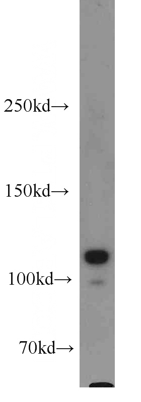
MCF7 cells were subjected to SDS PAGE followed by western blot with Catalog No:111705(HSPH1 antibody) at dilution of 1:1000
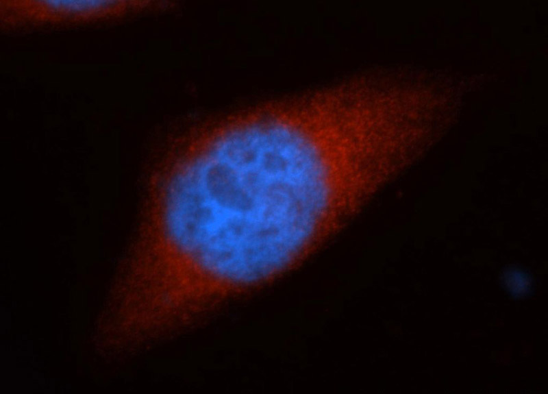
Immunofluorescent analysis of Hela cells, using HSPH1 antibody Catalog No:111705 at 1:50 dilution and Rhodamine-labeled goat anti-rabbit IgG (red). Blue pseudocolor = DAPI (fluorescent DNA dye).
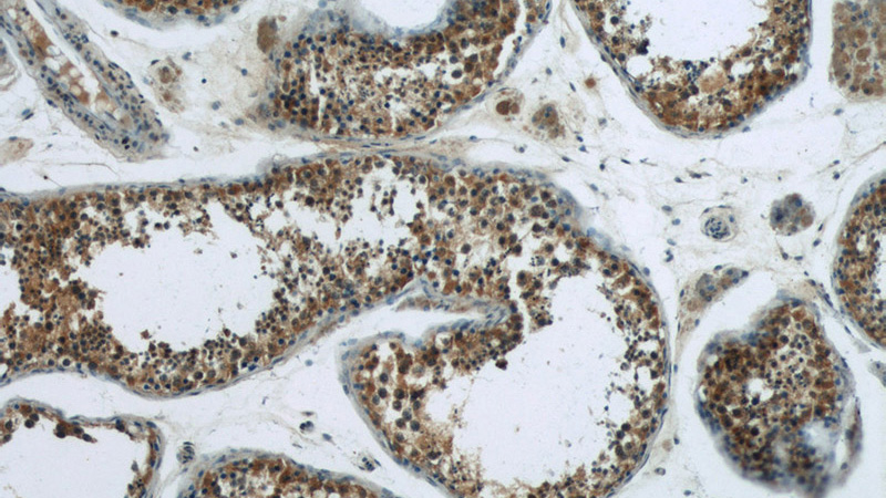
Immunohistochemistry of paraffin-embedded human testis tissue slide using Catalog No:111705(HSPH1 Antibody) at dilution of 1:50 (under 10x lens)
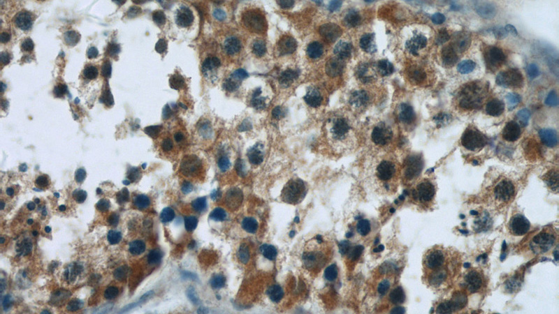
Immunohistochemistry of paraffin-embedded human testis tissue slide using Catalog No:111705(HSPH1 Antibody) at dilution of 1:50 (under 40x lens)
-
Background
HSP105, also known as HSP110 or HSPH1, belongs to the heat shock protein (HSP) family. Human HSP105 is a high-molecular-weight chaperone protein expressed at constitutively low levels as a cytoplasmic α-isoform and as an inducible nuclear β-isoform on exposure to various forms of stress. HSP105 is constitutively overexpressed in several solid tumors, including melanoma, breast, thyroid, and gastroenteric cancers, and exerts antiapoptotic functions. Recently HSP105 has been identified as a novel candidate biomarker of lymphoma aggressiveness. This antibody recognizes both HSP105α and HSP105β isoforms. Western blot analysis using this antibody detected a major band around 100-110kD in Jurkat cells.
-
References
- Yang L, Wang Y, Zhang Q. Identification of Hsf1 as a novel androgen receptor-regulated gene in mouse Sertoli cells. Molecular reproduction and development. 81(6):514-23. 2014.
Related Products / Services
Please note: All products are "FOR RESEARCH USE ONLY AND ARE NOT INTENDED FOR DIAGNOSTIC OR THERAPEUTIC USE"