
-
Product Name
Histone H1.0 antibody
- Documents
-
Description
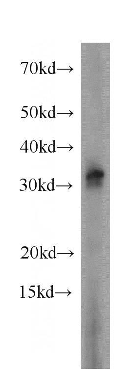
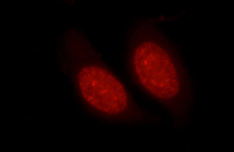
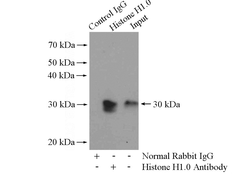
Histone H1.0 Rabbit Polyclonal antibody. Positive IF detected in HepG2 cells. Positive IP detected in A431 cells. Positive WB detected in A431 cells, HeLa cells, Jurkat cells, mouse spleen tissue, rat spleen tissue. Observed molecular weight by Western-blot: 32kd
-
Tested applications
ELISA, WB, IF, IP
-
Species reactivity
Human,Mouse,Rat; other species not tested.
-
Alternative names
H1 histone family antibody; member 0 antibody; H10 antibody; H1F0 antibody; H1FV antibody; Histone H1 antibody; Histone H1(0) antibody; Histone H1.0 antibody
-
Isotype
Rabbit IgG
-
Preparation
This antibody was obtained by immunization of Histone H1.0 recombinant protein (Accession Number: NM_005318). Purification method: Antigen affinity purified.
-
Clonality
Polyclonal
-
Formulation
PBS with 0.02% sodium azide and 50% glycerol pH 7.3.
-
Storage instructions
Store at -20℃. DO NOT ALIQUOT
-
Applications
Recommended Dilution:
WB: 1:500-1:5000
IP: 1:200-1:2000
IF: 1:20-1:200
-
Validations
A431 cells were subjected to SDS PAGE followed by western blot with Catalog No:111366(H1F0 antibody) at dilution of 1:500
Immunofluorescent analysis of HepG2 cells using Catalog No:111366(Histone H1.0 Antibody) at dilution of 1:50 and Rhodamine-Goat anti-Rabbit IgG
IP Result of anti-Histone H1.0 (IP:Catalog No:111366, 4ug; Detection:Catalog No:111366 1:500) with A431 cells lysate 2400ug.
-
Background
Histones are basic nuclear proteins that are responsible for the nucleosome structure of the chromosomal fiber in eukaryotes. Nucleosomes consist of approximately 146 bp of DNA wrapped around a histone octamer composed of pairs of each of the four core histones (H2A, H2B, H3, and H4). The chromatin fiber is further compacted through the interaction of a linker histone, H1, with the DNA between the nucleosomes to form higher order chromatin structures.Linker histones are involved in the formation of higher order structure in chromatin and the maintenance of overall chromatin compaction. The H1F0 histones are found in cells that are in terminal stages of differentiation or that have low rates of cell division.Histone H1.0 (H1F0,H1FV) is a linker histone that is widely expressed in many tissues and almost all vertebrates, unlike some other linker histones. The observed molecular weight of H1F0 is about 32 kDa.
-
References
- Poolman TM, Farrow SN, Matthews L, Loudon AS, Ray DW. Pin1 promotes GR transactivation by enhancing recruitment to target genes. Nucleic acids research. 41(18):8515-25. 2013.
- Tao M, Liu L, Shen M. Inflammatory stimuli promote growth and invasion of pancreatic cancer cells through NF-κB pathway dependent repression of PP2Ac. Cell cycle (Georgetown, Tex.). 15(3):381-93. 2016.
Related Products / Services
Please note: All products are "FOR RESEARCH USE ONLY AND ARE NOT INTENDED FOR DIAGNOSTIC OR THERAPEUTIC USE"