
-
Product Name
FUS/TLS antibody
- Documents
-
Description
FUS/TLS Rabbit Polyclonal antibody. Positive FC detected in HeLa cells. Positive IF detected in HepG2 cells. Positive IHC detected in human ovary tumor tissue, human brain tissue, human breast cancer tissue. Positive WB detected in HEK-293 cells, HEK-293, HeLa cells, HepG2 cells, HL-60 cells, human kidney tissue, Jurkat cells, K-562 cells, mouse brain tissue, SMMC-7721 cells. Positive IP detected in K-562 cells. Observed molecular weight by Western-blot: 73 kDa
-
Tested applications
ELISA, IF, IP, WB, IHC, FC
-
Species reactivity
Human,Mouse,Rat; other species not tested.
-
Alternative names
75 kDa DNA pairing protein antibody; FUS antibody; FUS1 antibody; FUS-CHOP antibody; hnRNP P2 antibody; Oncogene FUS antibody; Oncogene TLS antibody; POMp75 antibody; RNA binding protein FUS antibody; TLS antibody; TLS/CHOP antibody
- Immunogen
-
Isotype
Rabbit IgG
-
Preparation
This antibody was obtained by immunization of FUS/TLS recombinant protein (Accession Number: NM_004960). Purification method: Antigen affinity purified.
-
Clonality
Polyclonal
-
Formulation
PBS with 0.1% sodium azide and 50% glycerol pH 7.3.
-
Storage instructions
Store at -20℃. DO NOT ALIQUOT
-
Applications
Recommended Dilution:
WB: 1:1000-1:10000
IP: 1:500-1:5000
IHC: 1:20-1:200
IF: 1:10-1:100
-
Validations
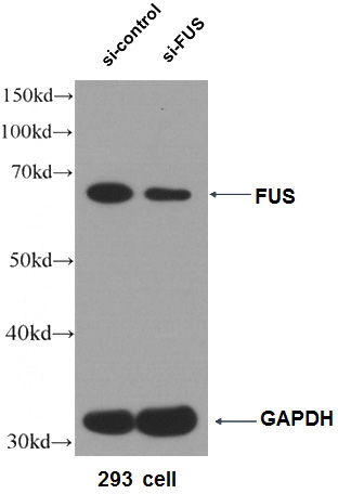
WB result of FUS antibody (Catalog No:110795, 1:5000) with si-Control and si-FUS transfected HEK 293 cells.
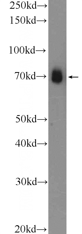
HEK-293 cells were subjected to SDS PAGE followed by western blot with Catalog No:110795(FUS/TLS Antibody) at dilution of 1:2000
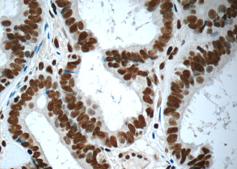
Immunohistochemistry of paraffin-embedded human ovary tumor tissue slide using Catalog No:110795(FUS/TLS Antibody) at dilution of 1:50 (under 40x lens)
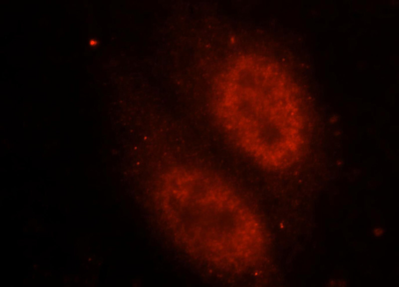
Immunofluorescent analysis of HepG2 cells, using FUS antibody Catalog No:110795 at 1:25 dilution and Rhodamine-labeled goat anti-rabbit IgG (red).
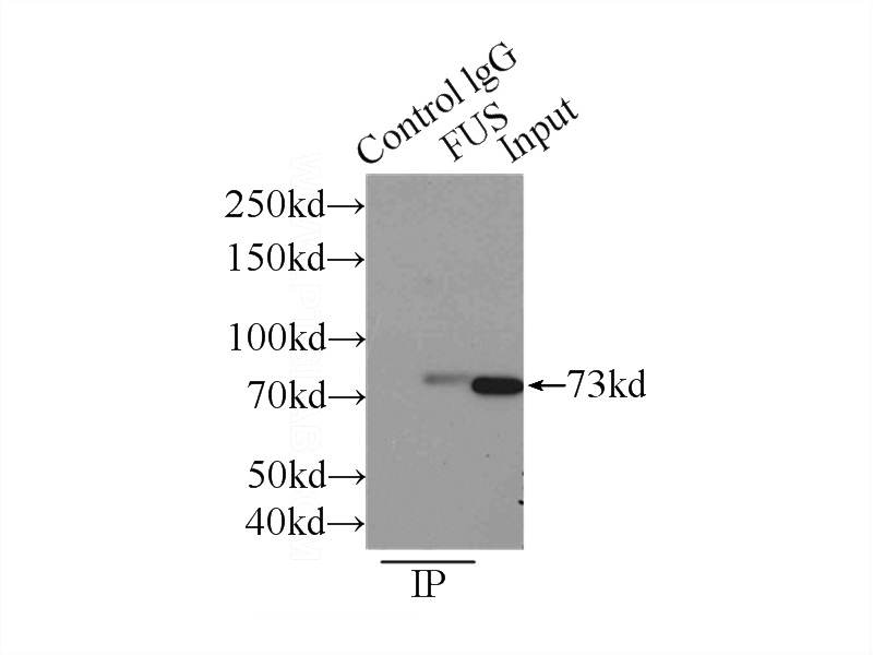
IP Result of anti-FUS (IP:Catalog No:110795, 3ug; Detection:Catalog No:110795 1:1000) with K-562 cells lysate 4000ug.
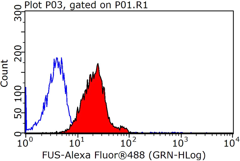
1X10^6 HeLa cells were stained with 0.2ug FUS/TLS antibody (Catalog No:110795, red) and control antibody (blue). Fixed with 90% MeOH blocked with 3% BSA (30 min). Alexa Fluor 488-congugated AffiniPure Goat Anti-Rabbit IgG(H+L) with dilution 1:1500.
-
Background
FUS (also named TLS and POMp75) belongs to the RRM TET family. FUS may play a role in the maintenance of genomic integrity; it binds both single-stranded and double-stranded DNA and promotes ATP-independent annealing of complementary single-stranded DNAs and D-loop formation in superhelical double-stranded DNA. FUS is also an RNA-binding protein, and its links to neurodegenerative disease proffer the intriguing possibility that altered RNA metabolism or RNA processing may underlie or contribute to neuron degeneration[PMID: 22640227]. FUS may be a cause of angiomatoid fibrous histiocytoma (AFH) and is implicated in certain forms of amyotrophic lateral sclerosis (ALS) and frontotemporal dementias (FTDs) such as frontotemporal lobar dementia with ubiquitin inclusions (FTLD-U)[PMID: 22640227]. This antibody is a rabbit polyclonal antibody raised against an internal region of human FUS.
-
References
- Rulten SL, Rotheray A, Green RL. PARP-1 dependent recruitment of the amyotrophic lateral sclerosis-associated protein FUS/TLS to sites of oxidative DNA damage. Nucleic acids research. 42(1):307-14. 2014.
- He J, Huang L, Qiu H. A new design of a lentiviral shRNA vector with inducible co-expression of ARGONAUTE 2 for enhancing gene silencing efficiency. Cell & bioscience. 5:67. 2015.
- Lim SM, Choi WJ, Oh KW. Directly converted patient-specific induced neurons mirror the neuropathology of FUS with disrupted nuclear localization in amyotrophic lateral sclerosis. Molecular neurodegeneration. 11(1):8. 2016.
- Kobayashi Z, Kawakami I, Arai T. Pathological features of FTLD-FUS in a Japanese population: analyses of nine cases. Journal of the neurological sciences. 335(1-2):89-95. 2013.
- Wong TH, Chiu WZ, Breedveld GJ. PRKAR1B mutation associated with a new neurodegenerative disorder with unique pathology. Brain : a journal of neurology. 137(Pt 5):1361-73. 2014.
- Acosta JR, Goldsbury C, Winnick C. Mutant human FUS Is ubiquitously mislocalized and generates persistent stress granules in primary cultured transgenic zebrafish cells. PloS one. 9(6):e90572. 2014.
- Smets K, Deconinck T, Baets J. Partial deletion of AFG3L2 causing spinocerebellar ataxia type 28. Neurology. 82(23):2092-100. 2014.
- Tibshirani M, Tradewell ML, Mattina KR. Cytoplasmic sequestration of FUS/TLS associated with ALS alters histone marks through loss of nuclear protein arginine methyltransferase 1. Human molecular genetics. 24(3):773-86. 2015.
Related Products / Services
Please note: All products are "FOR RESEARCH USE ONLY AND ARE NOT INTENDED FOR DIAGNOSTIC OR THERAPEUTIC USE"