

-
Product Name
FIS1 antibody
- Documents
-
Description
FIS1 Rabbit Polyclonal antibody. Positive WB detected in mouse brain tissue, A431 cells, COLO 320 cells, HEK-293 cells, HeLa cells, human heart tissue, human spleen tissue, mouse heart tissue, mouse thymus tissue, rat brain tissue, rat heart tissue. Positive IP detected in HeLa cells. Positive IF detected in Hepa1-6 cells. Positive IHC detected in human brain tissue, human prostate cancer tissue. Observed molecular weight by Western-blot: 17 kDa
-
Tested applications
ELISA, IHC, IF, IP, WB
-
Species reactivity
Human,Mouse,Rat; other species not tested.
-
Alternative names
CGI 135 antibody; FIS1 antibody; FIS1 homolog antibody; hFis1 antibody; TPR repeat protein 11 antibody; TTC11 antibody
-
Isotype
Rabbit IgG
-
Preparation
This antibody was obtained by immunization of FIS1 recombinant protein (Accession Number: NM_016068). Purification method: Antigen affinity purified.
-
Clonality
Polyclonal
-
Formulation
PBS with 0.1% sodium azide and 50% glycerol pH 7.3.
-
Storage instructions
Store at -20℃. DO NOT ALIQUOT
-
Applications
Recommended Dilution:
WB: 1:500-1:5000
IP: 1:200-1:2000
IHC: 1:10-1:100
IF: 1:10-1:100
-
Validations
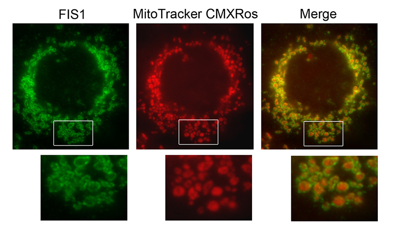
IF result of anti-FIS1(Catalog No:110667,1:100) with Hepa1-6 cell by Dr. Steven Eugene Smith. Mitochondrion outer membrane (Green) Stain.
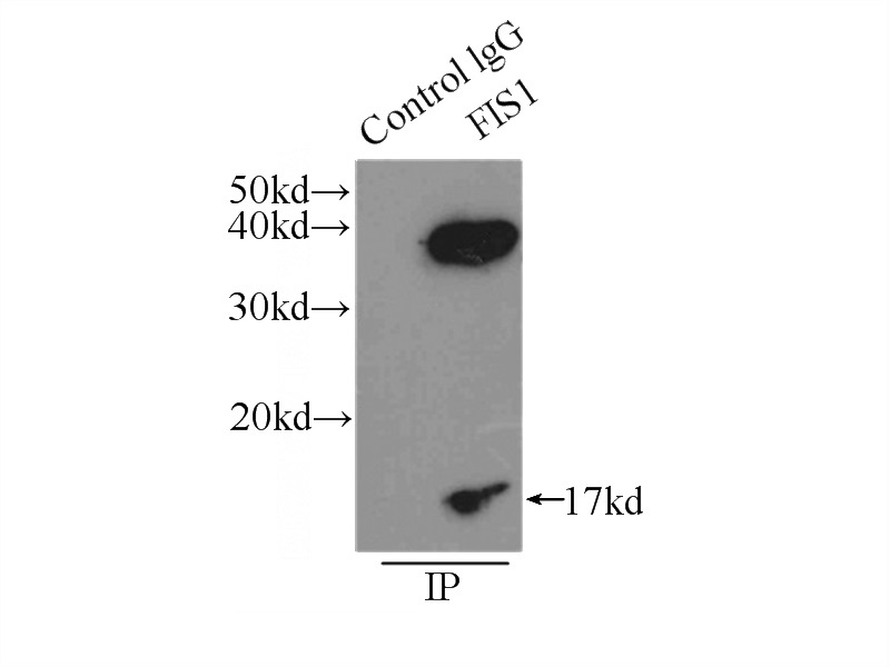
IP Result of anti-FIS1 (IP:Catalog No:110667, 3ug; Detection:Catalog No:110667 1:500) with HeLa cells lysate 3000ug.
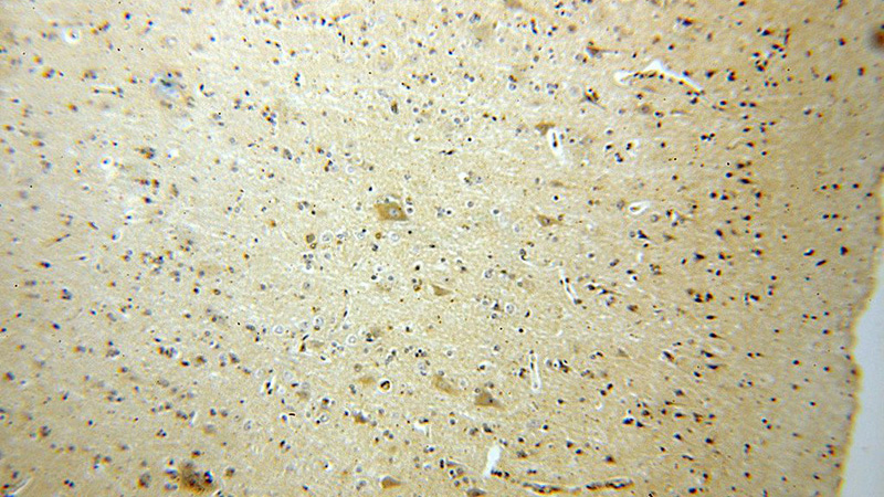
Immunohistochemical of paraffin-embedded human brain using Catalog No:110667(FIS1 antibody) at dilution of 1:25 (under 10x lens)
Immunohistochemical of paraffin-embedded human brain using Catalog No:110667(FIS1 antibody) at dilution of 1:25 (under 40x lens)
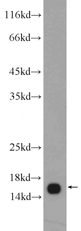
mouse brain tissue were subjected to SDS PAGE followed by western blot with Catalog No:110667(FIS1 Antibody) at dilution of 1:1000
-
Background
Fis1 (fission 1) is an integral mitochondrial outer membrane protein that participates in mitochondrial fission by interacting with dynamin-related protein 1 (Drp1). Excessive mitochondrial fission is associated with the pathology of a number of neurodegenerative or neurodevelopmental diseases. Increased expression of Fis1 has been found in Huntington’s disease (HD)-affected brain, Alzheimer’s disease (AD) patients, and autism spectrum disorder. This antibody was raised against the full-length of human Fis1 protein, and recognizes endogenous Fis1 protein in various lysates. (21257639, 21459773, 23333625)
-
References
- Wang X, Jiang W, Yan Y. RNA viruses promote activation of the NLRP3 inflammasome through a RIP1-RIP3-DRP1 signaling pathway. Nature immunology. 15(12):1126-33. 2014.
- Shen Q, Yamano K, Head BP. Mutations in Fis1 disrupt orderly disposal of defective mitochondria. Molecular biology of the cell. 25(1):145-59. 2014.
- Anand R, Wai T, Baker MJ. The i-AAA protease YME1L and OMA1 cleave OPA1 to balance mitochondrial fusion and fission. The Journal of cell biology. 204(6):919-29. 2014.
- Ruan Y, Li H, Zhang K, Jian F, Tang J, Song Z. Loss of Yme1L perturbates mitochondrial dynamics. Cell death & disease. 4:e896. 2013.
- Furuya N, Ikeda S, Sato S. PARK2/Parkin-mediated mitochondrial clearance contributes to proteasome activation during slow-twitch muscle atrophy via NFE2L1 nuclear translocation. Autophagy. 10(4):631-41. 2014.
- Manczak M, Reddy PH. Mitochondrial division inhibitor 1 protects against mutant huntingtin-induced abnormal mitochondrial dynamics and neuronal damage in Huntington's disease. Human molecular genetics. 24(25):7308-25. 2015.
- Sharoar MG, Shi Q, Ge Y. Dysfunctional tubular endoplasmic reticulum constitutes a pathological feature of Alzheimer's disease. Molecular psychiatry. 2015.
- Korwitz A, Merkwirth C, Richter-Dennerlein R. Loss of OMA1 delays neurodegeneration by preventing stress-induced OPA1 processing in mitochondria. The Journal of cell biology. 212(2):157-66. 2016.
Related Products / Services
Please note: All products are "FOR RESEARCH USE ONLY AND ARE NOT INTENDED FOR DIAGNOSTIC OR THERAPEUTIC USE"