
-
Product Name
ERp57/ERp60 antibody
- Documents
-
Description
ERp57/ERp60 Rabbit Polyclonal antibody. Positive IP detected in mouse liver tissue. Positive WB detected in L02 cells, A375 cells, mouse liver tissue, rat liver tissue. Positive IHC detected in human lung cancer tissue. Positive IF detected in HepG2 cells. Positive FC detected in HepG2 cells. Observed molecular weight by Western-blot: 57 kDa
-
Tested applications
ELISA, WB, IHC, IF, FC, IP
-
Species reactivity
Human,Mouse,Rat; other species not tested.
-
Alternative names
58 kDa microsomal protein antibody; Disulfide isomerase ER 60 antibody; ER protein 57 antibody; ER protein 60 antibody; ER60 antibody; ERp57 antibody; ERp60 antibody; ERp61 antibody; GRP57 antibody; GRP58 antibody; HsT17083 antibody; P58 antibody; PDIA3 antibody; PI PLC antibody; Protein disulfide isomerase A3 antibody
- Immunogen
-
Isotype
Rabbit IgG
-
Preparation
This antibody was obtained by immunization of ERp57/ERp60 recombinant protein (Accession Number: NM_005313). Purification method: Antigen affinity purified.
-
Clonality
Polyclonal
-
Formulation
PBS with 0.02% sodium azide and 50% glycerol pH 7.3.
-
Storage instructions
Store at -20℃. DO NOT ALIQUOT
-
Applications
Recommended Dilution:
WB: 1:500-1:5000
IP: 1:1000-1:10000
IHC: 1:20-1:200
IF: 1:20-1:200
-
Validations
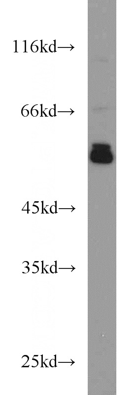
L02 cells were subjected to SDS PAGE followed by western blot with Catalog No:110374(PDIA3 antibody) at dilution of 1:1000
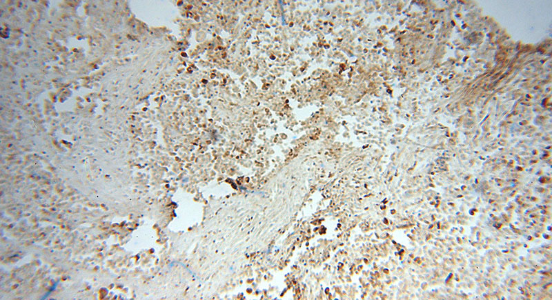
Immunohistochemical of paraffin-embedded human lung cancer using Catalog No:110374(PDIA3 antibody) at dilution of 1:100 (under 10x lens)
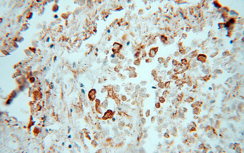
Immunohistochemical of paraffin-embedded human lung cancer using Catalog No:110374(PDIA3 antibody) at dilution of 1:100 (under 40x lens)
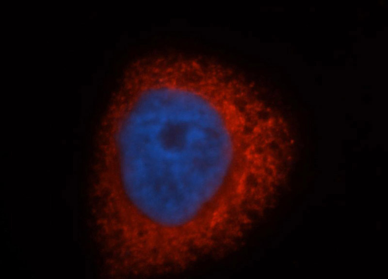
Immunofluorescent analysis of HepG2 cells, using PDIA3 antibody Catalog No:110374 at 1:50 dilution and Rhodamine-labeled goat anti-rabbit IgG (red). Blue pseudocolor = DAPI (fluorescent DNA dye).
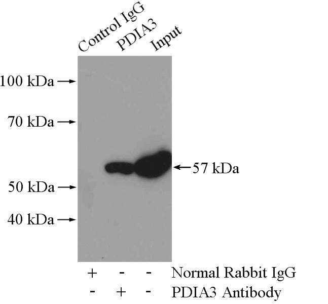
IP Result of anti-PDIA3 (IP:Catalog No:110374, 4ug; Detection:Catalog No:110374 1:2000) with mouse liver tissue lysate 4000ug.
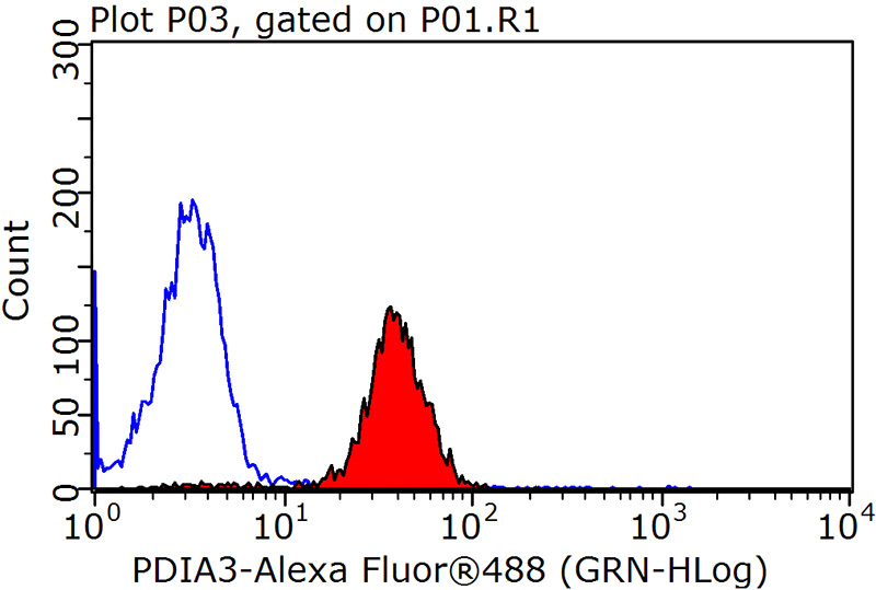
1X10^6 HepG2 cells were stained with 0.2ug PDIA3 antibody (Catalog No:110374, red) and control antibody (blue). Fixed with 90% MeOH blocked with 3% BSA (30 min). Alexa Fluor 488-congugated AffiniPure Goat Anti-Rabbit IgG(H+L) with dilution 1:1500.
-
Background
PDIA3, also named as P58, ER60, ERp57, ERp60, ERp61, GRP57, GRP58 and PI-PLC, is a member of the PDI family, participates in the oxidation, reduction, and isomerization of disulfide bonds for correct folding of secretory proteins before modification and transport in the endoplasmic reticulum. It is associated with apoptosis or inhibition of cancer cell growth. PDIA3 was once thought to be a phospholipase; however, it has been demonstrated that this protein actually has protein disulfide isomerase activity. It is thought that complexes of lectins and PDIA3 mediate protein folding by promoting formation of disulfide bonds in their glycoprotein substrates.
-
References
- Liu CI, Chen CC, Chen JC. Proteomic analysis of anti-tumor effects of 11-dehydrosinulariolide on CAL-27 cells. Marine drugs. 9(7):1254-72. 2011.
- Rocchiccioli S, Ucciferri N, Comelli L, Trivella MG, Citti L, Cecchettini A. Proteomics changes in adhesion molecules: a driving force for vascular smooth muscle cell phenotypic switch. Molecular bioSystems. 8(4):1052-9. 2012.
- Su CC, Su JH, Lin JJ. An investigation into the cytotoxic effects of 13-acetoxysarcocrassolide from the soft coral Sarcophyton crassocaule on bladder cancer cells. Marine drugs. 9(12):2622-42. 2011.
- Liu XM, Ding GL, Jiang Y. Down-regulation of S100A11, a calcium-binding protein, in human endometrium may cause reproductive failure. The Journal of clinical endocrinology and metabolism. 97(10):3672-83. 2012.
- Cheng KC, Huang HH, Hung CT. Proteomic analysis of the differences in orbital protein expression in thyroid orbitopathy. Graefe's archive for clinical and experimental ophthalmology = Albrecht von Graefes Archiv für klinische und experimentelle Ophthalmologie. 251(12):2777-87. 2013.
- Zhao Q, Feng Y, Jia X. Proteome analysis of hepatic non-parenchymal cells of immune liver fibrosis rats. Science China. Life sciences. 57(3):303-14. 2014.
- Chen K, Huang C, Yuan J, Cheng H, Zhou R. Long-term artificial selection reveals a role of TCTP in autophagy in mammalian cells. Molecular biology and evolution. 31(8):2194-211. 2014.
- Zhang X, Zuo X, Yang B. MicroRNA directly enhances mitochondrial translation during muscle differentiation. Cell. 158(3):607-19. 2014.
Related Products / Services
Please note: All products are "FOR RESEARCH USE ONLY AND ARE NOT INTENDED FOR DIAGNOSTIC OR THERAPEUTIC USE"