
-
Product Name
DAB2 antibody
- Documents
-
Description
DAB2 Rabbit Polyclonal antibody. Positive FC detected in HepG2 cells. Positive IF detected in HepG2 cells. Positive IP detected in HeLa cells. Positive WB detected in HeLa cells, MCF7 cells. Observed molecular weight by Western-blot: 96 kDa
-
Tested applications
ELISA, WB, IF, FC, IP
-
Species reactivity
Human,Mouse,Rat; other species not tested.
-
Alternative names
DAB2 antibody; Disabled homolog 2 antibody; DOC 2 antibody; DOC2 antibody; FLJ26626 antibody
-
Isotype
Rabbit IgG
-
Preparation
This antibody was obtained by immunization of DAB2 recombinant protein (Accession Number: NM_001343). Purification method: Antigen affinity purified.
-
Clonality
Polyclonal
-
Formulation
PBS with 0.1% sodium azide and 50% glycerol pH 7.3.
-
Storage instructions
Store at -20℃. DO NOT ALIQUOT
-
Applications
Recommended Dilution:
WB: 1:500-1:5000
IP: 1:1000-1:10000
IF: 1:20-1:200
-
Validations
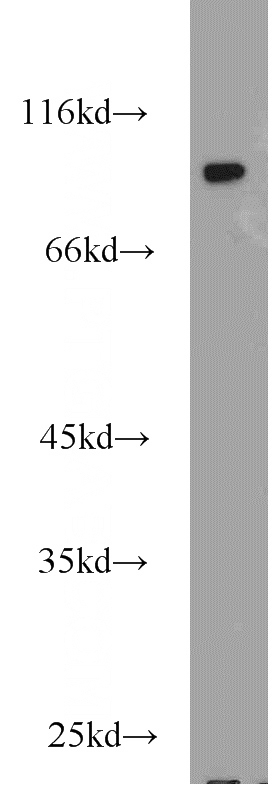
HeLa cells were subjected to SDS PAGE followed by western blot with Catalog No:109851(DAB2 antibody) at dilution of 1:1000
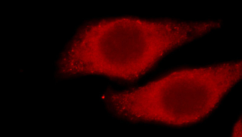
Immunofluorescent analysis of HepG2 cells, using DAB2 antibody Catalog No:109851 at 1:50 dilution and Rhodamine-labeled goat anti-rabbit IgG (red).
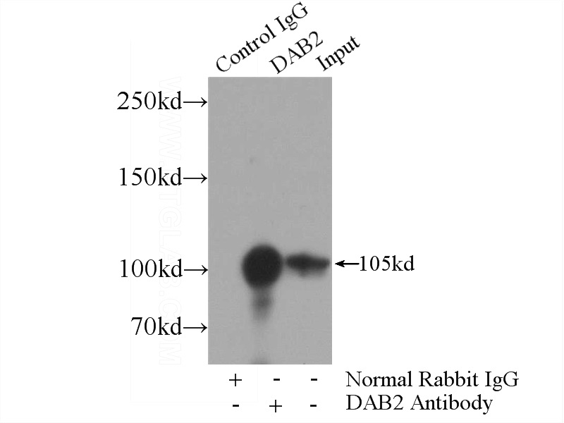
IP Result of anti-DAB2 (IP:Catalog No:109851, 4ug; Detection:Catalog No:109851 1:2000) with HeLa cells lysate 4000ug.
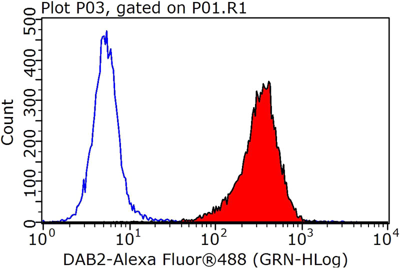
1X10^6 HepG2 cells were stained with 0.2ug DAB2 antibody (Catalog No:109851, red) and control antibody (blue). Fixed with 90% MeOH blocked with 3% BSA (30 min). Alexa Fluor 488-congugated AffiniPure Goat Anti-Rabbit IgG(H+L) with dilution 1:1000.
-
Background
DAB2 is a protein of 770 amino acid residues with a predicted molecular weight of 85.5kDa. This gene was initially named Doc2 (for Differentially expressed in Ovarian Cancer) and is distinct from the Doc2A and Doc2B genes (for double C2-like domains, alpha and beta). Human DAB2 has an overall 83% identify with the mouse p96 protein, a putative mitogen-responsive phosphoprotein; homology is strongest in the amino-terminal end of the protein in a region corresponding to the phosphotyrosine interaction domain (PID), and contains multiple SH3 binding motifs. Chromosomal localization by FISH showed that the DAB2 gene is located on 5p13. The expression of DAB2 is down-regulated or absent in all the carcinoma cell lines examined, including prostate and ovarian carcinoma cell lines. The N-terminal domain of DAB2 interacts with Dishevelled-3 (Dvl-3), a signaling mediator of the Wnt pathway. Ectopic expression of DAB2 attenuates canonical Wnt/catenin-mediated signaling, including accumulation of catenin and cyclin D1 induction. DAB2 suppresses both protein kinase C and peptide growth factor-elicited signal pathways via the Ras-mitogen-activated protein kinase pathway. The proline-rich domain of DAB2 also interacts with proteins containing SH3 domain, such as Src and Fgr. The binding of DAB2 to c-Src resultes in the inactivation of c-Src. All data suggest that DAB2 is a potent tumor suppressor in many cancer types.
-
References
- Mhawech-Fauceglia P, Kesterson J, Wang D. Expression and clinical significance of the transforming growth factor-β signalling pathway in endometrial cancer. Histopathology. 59(1):63-72. 2011.
Related Products / Services
Please note: All products are "FOR RESEARCH USE ONLY AND ARE NOT INTENDED FOR DIAGNOSTIC OR THERAPEUTIC USE"