
-
Product Name
CUL4A-Specific antibody
- Documents
-
Description
CUL4A-Specific Rabbit Polyclonal antibody. Positive WB detected in HeLa cells, HL-60 cells, Jurkat cells, mouse thymus tissue. Positive IF detected in HepG2 cells. Positive IHC detected in human liver cancer tissue. Observed molecular weight by Western-blot: 88 kDa,77 kDa
-
Tested applications
ELISA, IF, WB, IHC
-
Species reactivity
Human,Mouse,Rat; other species not tested.
-
Alternative names
CUL 4A antibody; CUL4A antibody; cullin 4A antibody
-
Isotype
Rabbit IgG
-
Preparation
This antibody was obtained by immunization of Peptide (Accession Number: NM_003589). Purification method: Antigen affinity purified.
-
Clonality
Polyclonal
-
Formulation
PBS with 0.02% sodium azide and 50% glycerol pH 7.3.
-
Storage instructions
Store at -20℃. DO NOT ALIQUOT
-
Applications
Recommended Dilution:
WB: 1:200-1:2000
IHC: 1:20-1:200
IF: 1:20-1:200
-
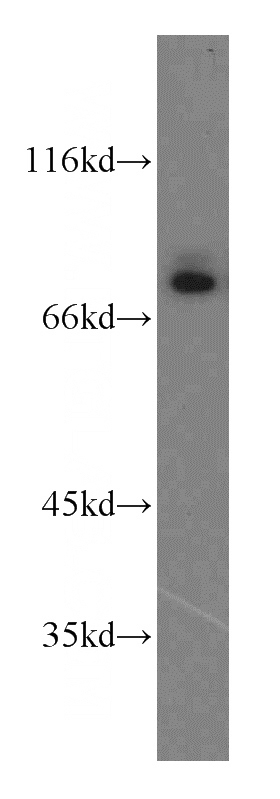
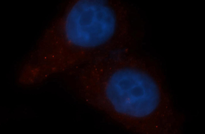
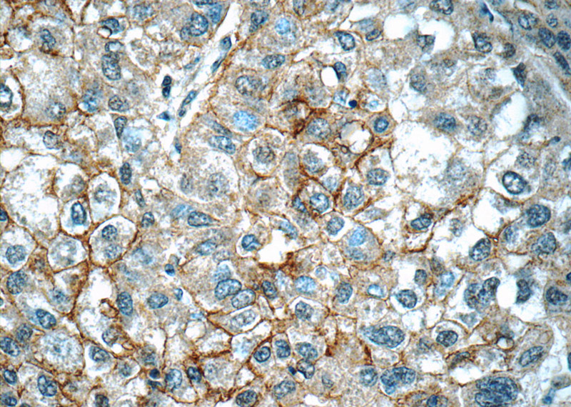
Validations
HeLa cells were subjected to SDS PAGE followed by western blot with Catalog No:109745(CUL4A-Specific antibody) at dilution of 1:500
Immunofluorescent analysis of HepG2 cells, using CUL4A antibody Catalog No: at 1:50 dilution and Rhodamine-labeled goat anti-rabbit IgG (red). Blue pseudocolor = DAPI (fluorescent DNA dye).
Immunohistochemistry of paraffin-embedded human liver cancer tissue slide using Catalog No:109745(CUL4A-Specific Antibody) at dilution of 1:50 (under 40x lens)
-
Background
Cullin proteins assemble a large number of RING E3 ubiquitin ligases, participating in the proteolysis through the ubiquitin-proteasome pathway. Two cullin 4 (CUL4) proteins, CUL4A (87 kDa) and CUL4B(104 kDa), have been identified. The two CUL4 sequences are 83% identical. They target certain proteins for degradation by binding protein DDB1 to form a CUL4-DDB1 ubiquitin ligase complex with DDB. They form two individual E3 ligases, DDB1-CUL4ADDB2 and DDB1-CUL4BDDB2 in this process. CUL4A appeared in both the nucleus and the cytosol, suggesting a more complex mechanism for entering the nucleus. CUL4B is localized in the nucleus and facilitates the transfer of DDB1 into the nucleus independently of DDB2. This antibody is specially against CUL4A,but not CUL4B.
-
References
- Antonioli M, Albiero F, Nazio F. AMBRA1 interplay with cullin E3 ubiquitin ligases regulates autophagy dynamics. Developmental cell. 31(6):734-46. 2014.
- Menon S, Chi H, Zhang H, Deng XW, Flavell RA, Wei N. COP9 signalosome subunit 8 is essential for peripheral T cell homeostasis and antigen receptor-induced entry into the cell cycle from quiescence. Nature immunology. 8(11):1236-45. 2007.
- Chen J, Shin JH, Zhao R. CSN6 drives carcinogenesis by positively regulating Myc stability. Nature communications. 5:5384. 2014.
Related Products / Services
Please note: All products are "FOR RESEARCH USE ONLY AND ARE NOT INTENDED FOR DIAGNOSTIC OR THERAPEUTIC USE"