
-
Product Name
COXIV antibody
- Documents
-
Description
COXIV Rabbit Polyclonal antibody. Positive WB detected in MCF7 cells, A375 cells, A431 cells, A549 cells, HEK-293 cells, HeLa cells, HepG2 cells, human brain tissue, human lung tissue, human skeletal muscle tissue, human skin tissue, human spleen tissue, Jurkat cells, L02 cells, mouse brain tissue, mouse heart tissue, mouse liver tissue, mouse skeletal muscle tissue, NIH/3T3 cells, rat brain tissue, rat liver tissue. Positive IP detected in mouse skeletal muscle tissue. Positive FC detected in HepG2 cells. Positive IHC detected in human prostate cancer tissue, human heart tissue, human pancreas tissue. Positive IF detected in HepG2 cells. Observed molecular weight by Western-blot: 17-18kd
-
Tested applications
ELISA, WB, IHC, IF, IP, FC
-
Species reactivity
Human,Mouse,Rat; other species not tested.
-
Alternative names
COX IV 1 antibody; COX4 antibody; COX4I1 antibody; COXIV antibody; CytOChrome c oxidase polypeptide IV antibody; CytOChrome c oxidase subunit 4 isoform 1 mitOChondrial antibody
-
Isotype
Rabbit IgG
-
Preparation
This antibody was obtained by immunization of COXIV recombinant protein (Accession Number: XM_047433623). Purification method: Antigen affinity purified.
-
Clonality
Polyclonal
-
Formulation
PBS with 0.1% sodium azide and 50% glycerol pH 7.3.
-
Storage instructions
Store at -20℃. DO NOT ALIQUOT
-
Applications
Recommended Dilution:
WB: 1:500-1:5000
IP: 1:500-1:5000
IHC: 1:20-1:200
IF: 1:20-1:200
-
Validations
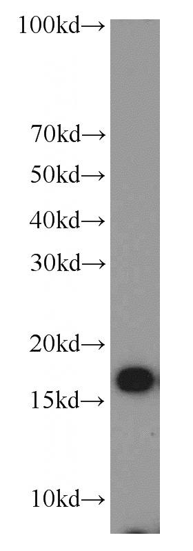
MCF7 cells were subjected to SDS PAGE followed by western blot with Catalog No:117310(COXIV antibody) at dilution of 1:1000
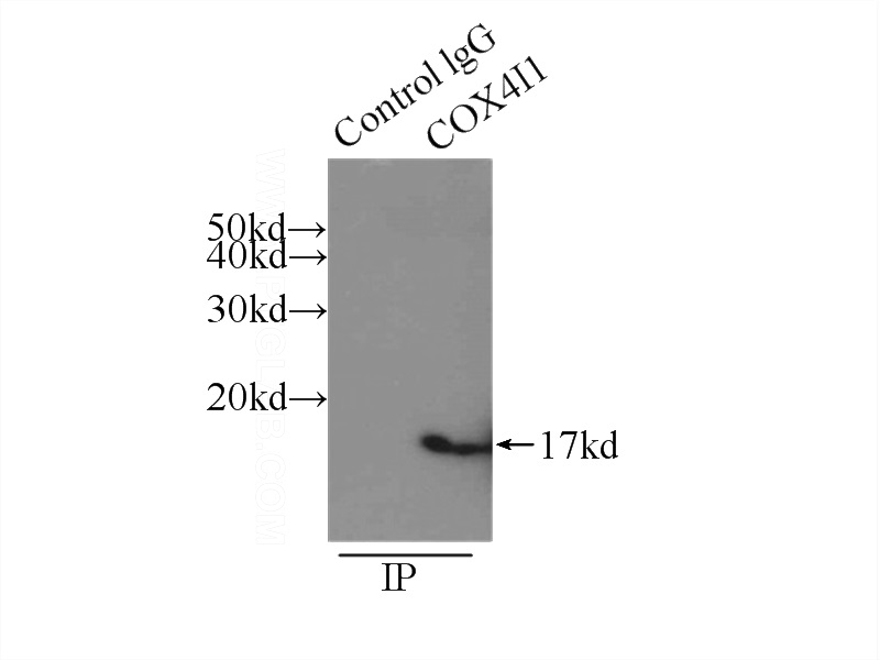
IP Result of anti-COXIV (IP:Catalog No:117310, 3ug; Detection:Catalog No:117310 1:1000) with mouse skeletal muscle tissue lysate 3500ug.
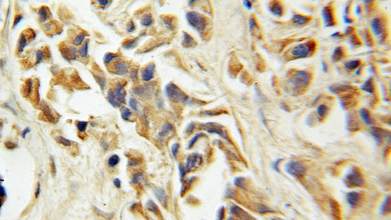
Immunohistochemical of paraffin-embedded human prostate cancer using Catalog No:117310(COXIV antibody) at dilution of 1:100 (under 10x lens)
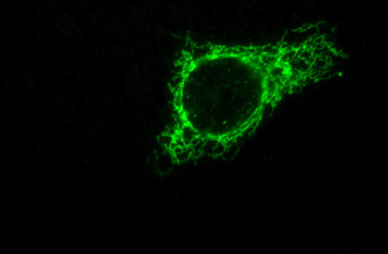
Immunofluorescent analysis of HepG2 cells, using COX4I1 antibody Catalog No:117310 at 1:50 dilution and FITC-labeled donkey anti-rabbit IgG(green).
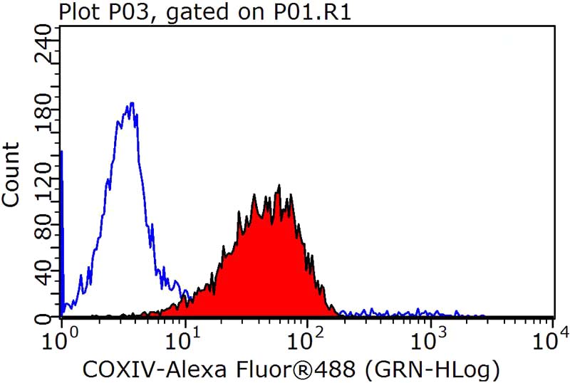
1X10^6 HepG2 cells were stained with .2ug COXIV antibody (Catalog No:117310, red) and control antibody (blue). Fixed with 90% MeOH blocked with 3% BSA (30 min). Alexa Fluor 488-congugated AffiniPure Goat Anti-Rabbit IgG(H+L) with dilution 1:1000.
-
Background
COX4I1, also named as COX4 and COXIV-1, belongs to the cytochrome c oxidase IV family. It is one of the nuclear-coded polypeptide chains of cytochrome c oxidase, the terminal oxidase in mitochondrial electron transport. COX4I1 is a marker for mitochondria. It has two isoforms (isoform 1 and 2). Isoform 1(COX4I1) is ubiquitously expressed and isoform 2 is highly expressed in lung tissues. COX4I1 is commonly used as a loading control. This antibody was generated against full length COX4I1 protein and cross reacts with COX4I2.
-
References
- Chen W, Hou J, Yin Y. alpha-Bisabolol induces dose- and time-dependent apoptosis in HepG2 cells via a Fas- and mitochondrial-related pathway, involves p53 and NFkappaB. Biochemical pharmacology. 80(2):247-54. 2010.
- Du J, Ma M, Zhao Q. Mitochondrial bioenergetic deficits in the hippocampi of rats with chronic ischemia-induced vascular dementia. Neuroscience. 231:345-52. 2013.
- Zhao L, Liu X, Liang J. Phosphorylation of p38 MAPK mediates hypoxic preconditioning-induced neuroprotection against cerebral ischemic injury via mitochondria translocation of Bcl-xL in mice. Brain research. 1503:78-88. 2013.
- Song T, Liang F, Zhang Z, Liu Y, Sheng H, Xie M. S1 kills MCF-7/ADR cells more than MCF-7 cells: A protective mechanism of endoplasmic reticulum stress. Biomedicine & pharmacotherapy = Biomédecine & pharmacothérapie. 67(8):731-6. 2013.
- Zhang H, Guo S, Zhang L. Treatment with carnosine reduces hypoxia-ischemia brain damage in a neonatal rat model. European journal of pharmacology. 727:174-80. 2014.
- Chen YY, Liu SL, Hu DP, Xing YQ, Shen Y. N -methyl- N -nitrosourea-induced retinal degeneration in mice. Experimental eye research. 121:102-13. 2014.
- Sun S, Han Y, Liu J. Trichostatin A targets the mitochondrial respiratory chain, increasing mitochondrial reactive oxygen species production to trigger apoptosis in human breast cancer cells. PloS one. 9(3):e91610. 2014.
- Aras S, Bai M, Lee I, Springett R, Hüttemann M, Grossman LI. MNRR1 (formerly CHCHD2) is a bi-organellar regulator of mitochondrial metabolism. Mitochondrion. 20:43-51. 2015.
Related Products / Services
Please note: All products are "FOR RESEARCH USE ONLY AND ARE NOT INTENDED FOR DIAGNOSTIC OR THERAPEUTIC USE"