
-
Product Name
CISD1 antibody
- Documents
-
Description
CISD1 Rabbit Polyclonal antibody. Positive FC detected in HeLa cells. Positive IF detected in Hela cells. Positive IHC detected in human heart tissue, human brain tissue, human kidney tissue, human liver tissue, human ovary tissue, human placenta tissue, human spleen tissue, human testis tissue. Positive IP detected in HepG2 cells. Positive WB detected in mouse skeletal muscle tissue, HepG2 cells, human heart tissue, human liver tissue, human skeletal muscle tissue, mouse brain tissue, mouse heart tissue, mouse kidney tissue. Observed molecular weight by Western-blot: 17 kDa
-
Tested applications
ELISA, WB, IHC, IF, IP, FC
-
Species reactivity
Human, Mouse, Zebrafish, Rat; other species not tested.
-
Alternative names
C10orf70 antibody; CDGSH iron sulfur domain 1 antibody; CISD1 antibody; MDS029 antibody; mitoNEET antibody; Mnt antibody; ZCD1 antibody
-
Isotype
Rabbit IgG
-
Preparation
This antibody was obtained by immunization of CISD1 recombinant protein (Accession Number: NM_018464). Purification method: Antigen affinity purified.
-
Clonality
Polyclonal
-
Formulation
PBS with 0.02% sodium azide and 50% glycerol pH 7.3.
-
Storage instructions
Store at -20℃. DO NOT ALIQUOT
-
Applications
Recommended Dilution:
WB: 1:1000-1:10000
IP: 1:1000-1:10000
IHC: 1:20-1:200
IF: 1:10-1:100
-
Validations
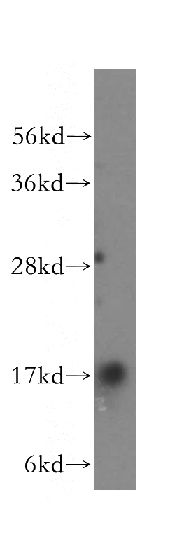
mouse skeletal muscle tissue were subjected to SDS PAGE followed by western blot with Catalog No:109321(mitoNEET,CISD1 antibody) at dilution of 1:4000
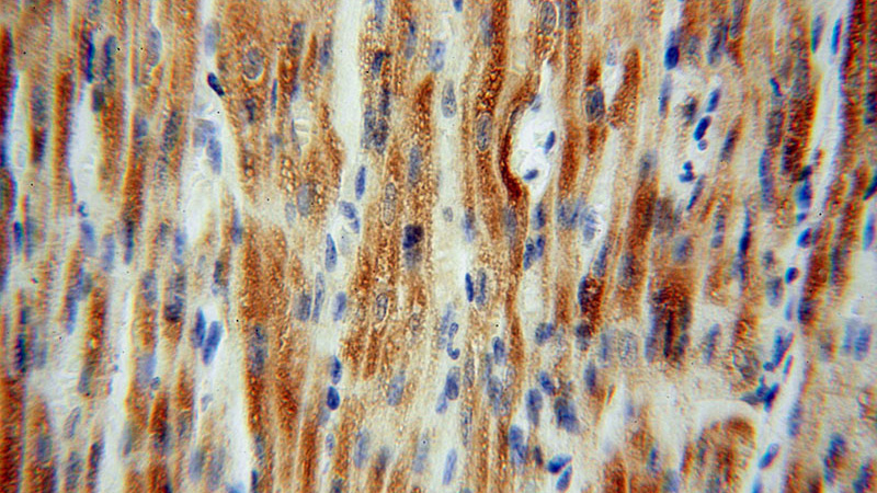
Immunohistochemical of paraffin-embedded human heart using Catalog No:109321(mitoNEET,CISD1 antibody) at dilution of 1:100 (under 40x lens)
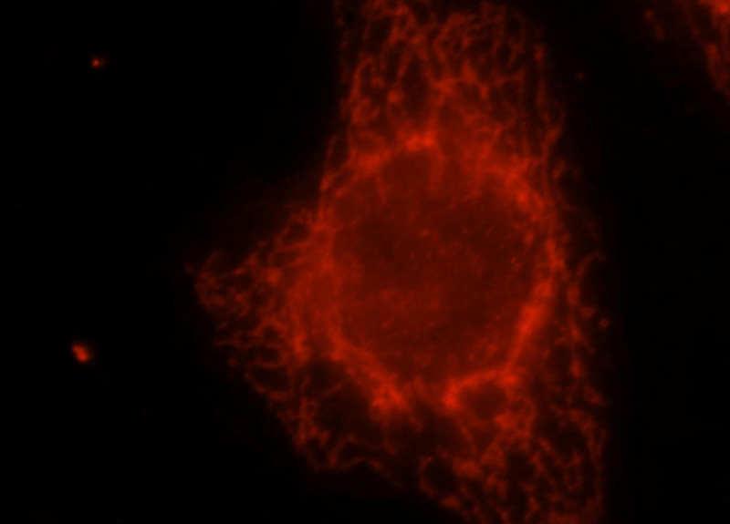
Immunofluorescent analysis of Hela cells, using CISD1 antibody Catalog No:109321 at 1:25 dilution and Rhodamine-labeled goat anti-rabbit IgG (red).
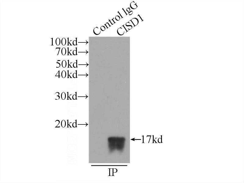
IP Result of anti-mitoNEET,CISD1 (IP:Catalog No:109321, 3ug; Detection:Catalog No:109321 1:2000) with HepG2 cells lysate 600ug.
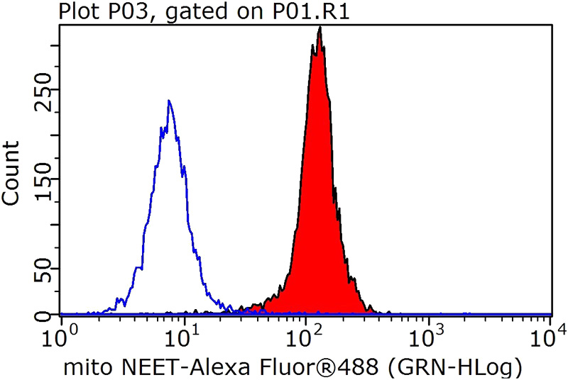
1X10^6 HeLa cells were stained with .05ug mitoNEET,CISD1 antibody (Catalog No:109321, red) and control antibody (blue). Fixed with 90% MeOH blocked with 3% BSA (30 min). Alexa Fluor 488-Goat anti-Rabbit IgG with dilution 1:100.
-
Background
MitoNEET, also named CISD1, belongs to a previously uncharacterized ancient family of proteins for which the hallmark is the presence of a unique 39 amino acid CDGSH domain. It is a single-pass type III membrane protein, located in mitochondrion outer membrane and may play a role in regulating maximal capacity for electron transport and oxidative phosphorylation. MitoNEET is a recently identified drug target for a commonly prescribed diabetes drug, Pioglitazone. This antibody recognizing MitoNEET (calculated 12 kDa) as a 17 kDa protein may be due to its posttranslational modification or metal binding activity.
-
References
- Lazarou M, Narendra DP, Jin SM, Tekle E, Banerjee S, Youle RJ. PINK1 drives Parkin self-association and HECT-like E3 activity upstream of mitochondrial binding. The Journal of cell biology. 200(2):163-72. 2013.
- Narendra DP, Wang C, Youle RJ, Walker JE. PINK1 rendered temperature sensitive by disease-associated and engineered mutations. Human molecular genetics. 22(13):2572-89. 2013.
- Fajardo VA, McMeekin L, Basic A, Lamb GD, Murphy RM, LeBlanc PJ. Isolation of sarcolemmal plasma membranes by mechanically skinning rat skeletal muscle fibers for phospholipid analysis. Lipids. 48(4):421-30. 2013.
- Shabalina IG, Landreh L, Edgar D. Leydig cell steroidogenesis unexpectedly escapes mitochondrial dysfunction in prematurely aging mice. FASEB journal : official publication of the Federation of American Societies for Experimental Biology. 29(8):3274-86. 2015.
- Ordureau A, Heo JM, Duda DM. Defining roles of PARKIN and ubiquitin phosphorylation by PINK1 in mitochondrial quality control using a ubiquitin replacement strategy. Proceedings of the National Academy of Sciences of the United States of America. 112(21):6637-42. 2015.
- Kazlauskaite A, Martínez-Torres RJ, Wilkie S. Binding to serine 65-phosphorylated ubiquitin primes Parkin for optimal PINK1-dependent phosphorylation and activation. EMBO reports. 16(8):939-54. 2015.
- Ordureau A, Sarraf SA, Duda DM. Quantitative proteomics reveal a feedforward mechanism for mitochondrial PARKIN translocation and ubiquitin chain synthesis. Molecular cell. 56(3):360-75. 2014.
- Lai YC, Kondapalli C, Lehneck R. Phosphoproteomic screening identifies Rab GTPases as novel downstream targets of PINK1. The EMBO journal. 34(22):2840-61. 2015.
Related Products / Services
Please note: All products are "FOR RESEARCH USE ONLY AND ARE NOT INTENDED FOR DIAGNOSTIC OR THERAPEUTIC USE"