
-
Product Name
CDA1 antibody
- Documents
-
Description
CDA1 Rabbit Polyclonal antibody. Positive IHC detected in human lung cancer tissue. Positive IF detected in Hela cells. Positive IP detected in HEK-293 cells. Positive WB detected in HeLa cells, DU 145 cells, HEK-293 cells, MCF7 cells. Observed molecular weight by Western-blot: 120 kDa
-
Tested applications
ELISA, IHC, IF, IP, WB
-
Species reactivity
Human; other species not tested.
-
Alternative names
CDA1 antibody; Cell division autoantigen 1 antibody; CINAP antibody; CTCL antibody; DENTT antibody; NP79 antibody; Nuclear protein of 79 kDa antibody; TSPX antibody; TSPYL2 antibody
-
Isotype
Rabbit IgG
-
Preparation
This antibody was obtained by immunization of CDA1 recombinant protein (Accession Number: NM_022117). Purification method: Antigen affinity purified.
-
Clonality
Polyclonal
-
Formulation
PBS with 0.1% sodium azide and 50% glycerol pH 7.3.
-
Storage instructions
Store at -20℃. DO NOT ALIQUOT
-
Applications
Recommended Dilution:
WB: 1:500-1:5000
IP: 1:500-1:5000
IHC: 1:20-1:200
IF: 1:20-1:200
-
Validations
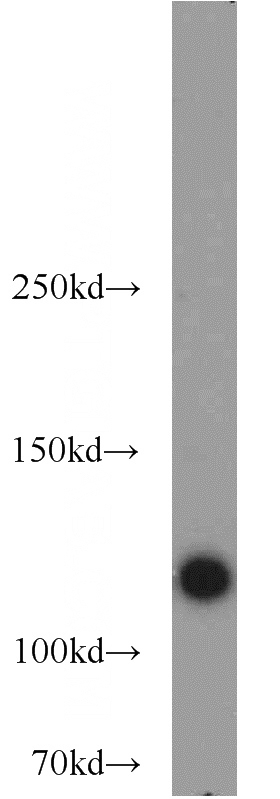
HeLa cells were subjected to SDS PAGE followed by western blot with Catalog No:109089(TSPYL2,TSPX,NP79 antibody) at dilution of 1:1000
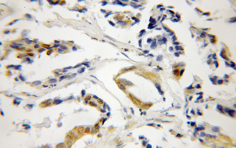
Immunohistochemical of paraffin-embedded human lung cancer using Catalog No:109089(TSPYL2,TSPX,NP79 antibody) at dilution of 1:50 (under 40x lens)
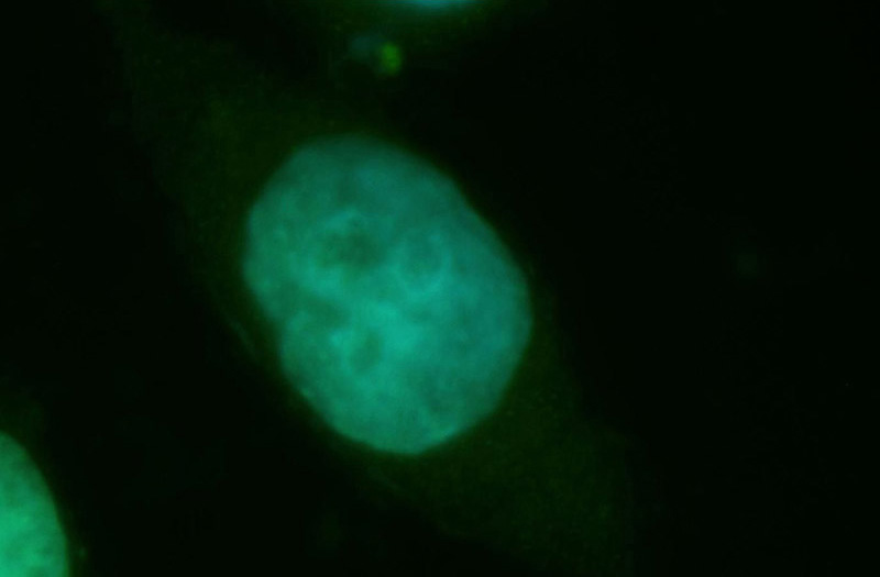
Immunofluorescent analysis of Hela cells, using TSPYL2 antibody Catalog No:109089 at 1:50 dilution and FITC-labeled donkey anti-rabbit IgG (green). Blue pseudocolor = DAPI (fluorescent DNA dye).
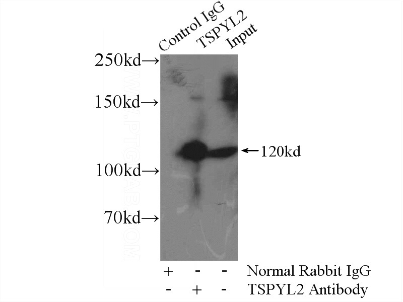
IP Result of anti-TSPYL2,TSPX,NP79 (IP:Catalog No:109089, 4ug; Detection:Catalog No:109089 1:1000) with HEK-293 cells lysate 1000ug.
-
Background
TSPYL2 (also known as CINAP, CDA1, TSPX or DENTT) is a new member of the nucleosome assembly protein superfamily. TSPYL2 binds histones and facilitates nucleosome assembly. TSPYL2 is expressed in various tissues, highly in the pituitary gland and moderately in the adrenals, brain, testis, and ovary. Immunohistochemical staining analysis for TSPYL2 showed differential cytoplasmic and nuclear staining patterns in several cell types. Downregulated expression of TSPYL2 has been observed in several tumors, which suggests its role as a tumor suppressor. Although it is predicted that TSPYL2 has a molecular mass of 79.43 kDa, it is found that mammalian TSPYL2 appears at a size of 120 kDa by western blot analysis. The abundant acidic amino acid regions in TSPYL2 may cause its aberrant migration. In addition, the TSPYL2 protein is unstable and sensitive to proteasomal degradation.
-
References
- Epping MT, Lunardi A, Nachmani D. TSPYL2 is an essential component of the REST/NRSF transcriptional complex for TGFβ signaling activation. Cell death and differentiation. 22(8):1353-62. 2015.
- Conrad S, Azizi H, Hatami M. Expression of Genes Related to Germ Cell Lineage and Pluripotency in Single Cells and Colonies of Human Adult Germ Stem Cells. Stem cells international. 2016:8582526. 2016.
- Eyler CE, Wu Q, Yan K. Glioma stem cell proliferation and tumor growth are promoted by nitric oxide synthase-2. Cell. 146(1):53-66. 2011.
- Kido T, Ou JH, Lau YF. The X-linked tumor suppressor TSPX interacts and promotes degradation of the hepatitis B viral protein HBx via the proteasome pathway. PloS one. 6(7):e22979. 2011.
Related Products / Services
Please note: All products are "FOR RESEARCH USE ONLY AND ARE NOT INTENDED FOR DIAGNOSTIC OR THERAPEUTIC USE"