

-
Product Name
ATP5A1 antibody
- Documents
-
Description
ATP5A1 Mouse Monoclonal antibody. Positive IHC detected in human heart tissue, human liver tissue. Positive IF detected in Hela cells. Positive FC detected in HeLa cells. Positive WB detected in multi-cells, HeLa cells, MCF7 cells. Positive IP detected in mouse heart tissue. Observed molecular weight by Western-blot: 50 kDa
-
Tested applications
ELISA, IHC, IF, WB, FC, IP
-
Species reactivity
Human, Mouse, Rat, Monkey; other species not tested.
-
Alternative names
ATP5A antibody; ATP5A1 antibody; ATP5AL2 antibody; ATPM antibody; hATP1 antibody; MOM2 antibody; OMR antibody; ORM antibody
-
Isotype
Mouse IgG2b
-
Preparation
This antibody was obtained by immunization of ATP5A1 recombinant protein (Accession Number: NM_004046). Purification method: Protein A purified.
-
Clonality
Monoclonal
-
Formulation
PBS with 0.02% sodium azide and 50% glycerol pH 7.3.
-
Storage instructions
Store at -20℃. DO NOT ALIQUOT
-
Applications
Recommended Dilution:
WB: 1:500-1:5000
IP: 1:200-1:2000
IHC: 1:20-1:200
IF: 1:20-1:200
-
Validations
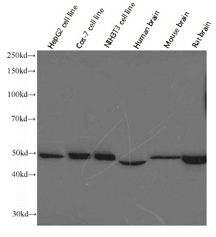
Western blot analysis of ATP5A1 in various tissues and cell lines using Proteintech antibody Catalog No:107071 at the dilution of 1:1000.
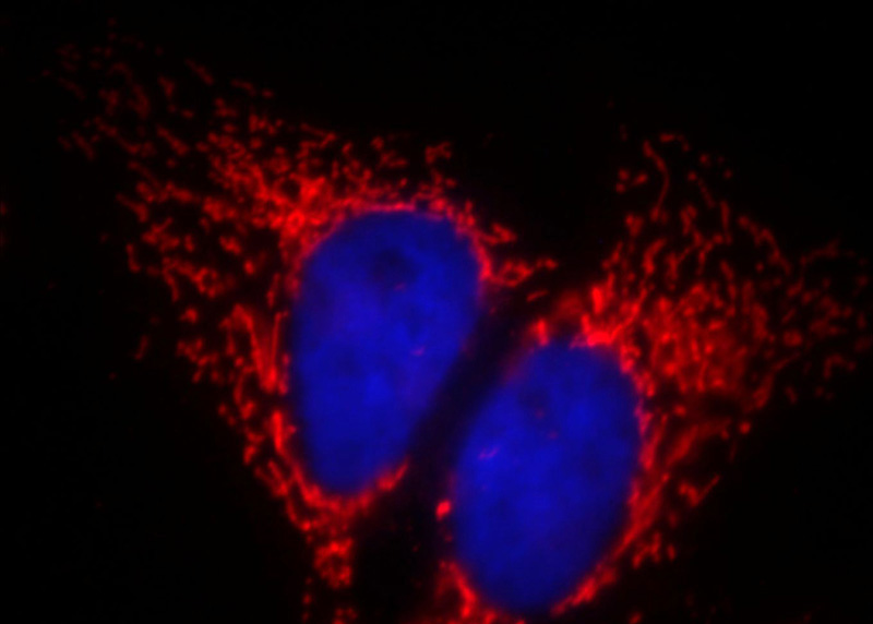
Immunofluorescent analysis of Hela cells, using ATP5A1 antibody Catalog No: at 1:50 dilution and Rhodamine-labeled goat anti-mouse IgG (red). Blue pseudocolor = DAPI (fluorescent DNA dye).
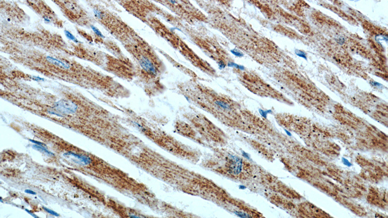
Immunohistochemical of paraffin-embedded human heart using Catalog No:107071(ATP5A1 antibody) at dilution of 1:50 (under 10x lens)
Immunohistochemical of paraffin-embedded human heart using Catalog No:107071(ATP5A1 antibody) at dilution of 1:50 (under 40x lens)
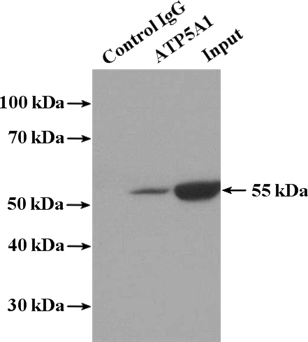
IP Result of anti-ATP5A1 (IP:Catalog No:107071, 5ug; Detection:Catalog No:107071 1:500) with mouse heart tissue lysate 4000ug.
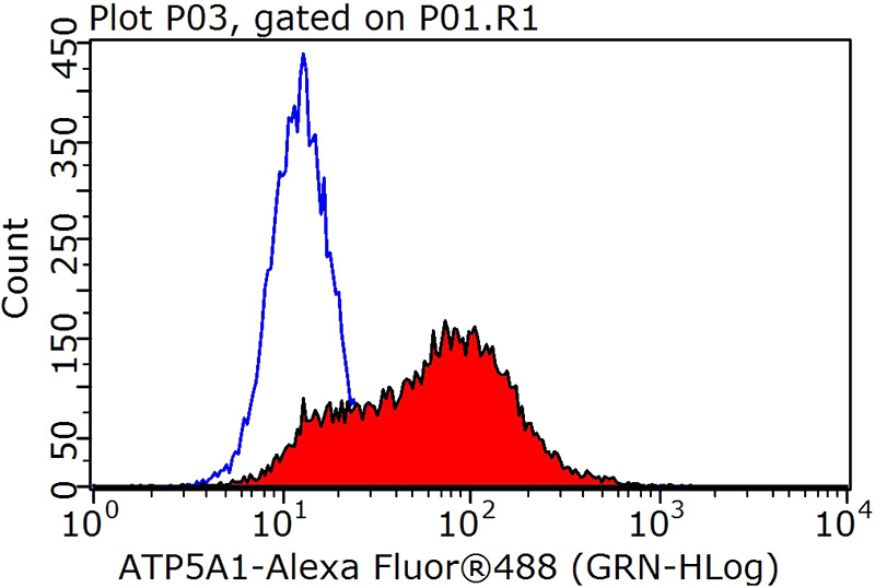
1X10^6 HeLa cells were stained with 0.2ug ATP5A1 antibody (Catalog No:107071, red) and control antibody (blue). Fixed with 90% MeOH blocked with 3% BSA (30 min). Alexa Fluor 488-congugated AffiniPure Goat Anti-Mouse IgG(H+L) with dilution 1:1000.
-
Background
The ATP5A1 gene encodes the α subunit of mitochondrial ATP synthase which produces ATP from ADP in the presence of a proton gradient across the membrane. The mitochondrial ATP synthase, also known as Complex V or F1F0 ATP synthase, is a multi-subunit enzyme complex consisting of two functional domains, the F1-containing the catalytic core and the Fo- containing the membrane proton channel. F0 domain has 10 subunits: a,b, c, d, e, f, g, OSCP, A6L, and F6. F1 is composed of subunits α, β, γ, δ, ε, and a loosely attached inhibitor protein IF1. Recently defect in ATP5A1 has been linked to the fatal neonatal mitochondrial encephalopathy. ATP5A1 is localized in the mitochondria and anti-ATP5A1 can be used as the loading control for mitochondrial or Complex V proteins. This antibody recognizes the endogenous ATP5A1 protein in lysates from various cell lines and tissues.
-
References
- Du Y, Meng Y, Zhu J. Quantitative proteomic study of myocardial mitochondria in urea transporter B knockout mice. Proteomics. 14(17-18):2072-83. 2014.
Related Products / Services
Please note: All products are "FOR RESEARCH USE ONLY AND ARE NOT INTENDED FOR DIAGNOSTIC OR THERAPEUTIC USE"