
-
Product Name
Anti-GST3/GST pi (9D4) Mouse antibody
- Documents
-
Description
GST3/GST pi (9D4) Mouse monoclonal antibody
-
Tested applications
WB, IHC-P, ICC/IF, FC
-
Species reactivity
Human
-
Isotype
Mouse IgG1
-
Preparation
Antigen: Purified recombinant fragment of human GSTP1 expressed in E. Coli.
-
Clonality
Monoclonal
-
Formulation
Ascitic fluid containing 0.03% sodium azide.
-
Storage instructions
Store at 4°C short term. Store at -20°C long term. Avoid freeze / thaw cycle.
-
Applications
WB: 1/500 - 1/2000
IHC: 1/200 - 1/1000
ICC: 1/200 - 1/1000
FC: 1/200 - 1/400
ELISA: 1/10000
-
Validations
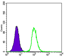
Flow cytometric analysis of K562 cells using GSTP1 mouse mAb (green) and negative control (purple).
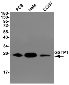
Western blot detection of GSTP1 in PC3,Hela,COS7 cell lysates using GSTP1 (9D4) Mouse mAb(1:1000 diluted).Predicted band size:23KDa.Observed band size:23KDa.
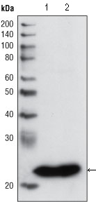
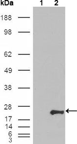
Western blot analysis using GSTP1 mouse mAb against PC3 cell lysate (1) and human cerebellum tissue lysate (2).
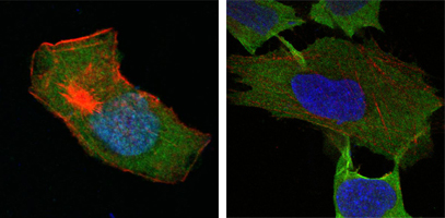
Confocal immunofluorescence analysis of HepG2 (left) and L-02 (right) cells using GSTP1 mouse mAb (green). Red: Actin filaments have been labeled with DY-554 phalloidin. Blue: DRAQ5 fluorescent DNA dye.
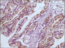
Immunohistochemical analysis of paraffin-embedded human prostate tissues using GSTP1 mouse mAb with DAB staining.
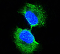
Confocal immunofluorescence analysis of PC-3 cells using GSTP1 mouse mAb (green). Blue: DRAQ5 fluorescent DNA dye.
Western blot analysis using GSTP1 mouse mAb against HEK293T cells transfected with the pCMV6-ENTRY control (1) and pCMV6-ENTRY GSTP1 cDNA (2).
-
Background
Swiss-Prot Acc.P09211.GSTP1 (glutathione-S-transferase, pi 1), also called GST3/DFN7, is a family of enzymes that play an important role in detoxification by catalyzing the conjugation of many hydrophobic and electrophilic compounds with reduced glutathione. GSTP1 act like a tumor suppressor gene, which when inactivated leads to tumor growth, and the -class glutathione S-transferase is commonly inactivated by somatic CpGisland hypermethylation in cancers of the prostate, liver, and breast. Methylation of regulatory sequences at the GSTP1 gene locus is found in the vast majority (>90%) of prostate carcinomas and is associated with transcriptional down-regulation.
Related Products / Services
Please note: All products are "FOR RESEARCH USE ONLY AND ARE NOT INTENDED FOR DIAGNOSTIC OR THERAPEUTIC USE"