
-
Product Name
AKT1 antibody
- Documents
-
Description
AKT1 Mouse Monoclonal antibody. Positive IP detected in mouse brain tissue. Positive WB detected in HeLa cells, rat liver tissue. Positive IHC detected in human cervical cancer tissue, human breast cancer tissue. Positive IF detected in MCF-7 cells. Positive FC detected in MCF-7 cells. Observed molecular weight by Western-blot: 62 kDa
-
Tested applications
ELISA, WB, IF, IHC, FC, IP
-
Species reactivity
Human,Mouse, Rat; other species not tested.
-
Alternative names
AKT antibody; AKT1 antibody; PKB antibody; PKB ALPHA antibody; PRKBA antibody; Protein kinase B antibody; Proto oncogene c Akt antibody; RAC antibody; RAC ALPHA antibody; RAC PK alpha antibody
-
Isotype
Mouse IgG1
-
Preparation
This antibody was obtained by immunization of AKT1 recombinant protein (Accession Number: NM_001382430). Purification method: .
-
Clonality
Monoclonal
-
Formulation
PBS with 0.02% sodium azide and 50% glycerol pH 7.3.
-
Storage instructions
Store at -20℃. DO NOT ALIQUOT
-
Applications
Recommended Dilution:
WB: 1:500-1:5000
IP: 1:200-1:2000
IHC: 1:20-1:200
IF: 1:20-1:200
-
Validations
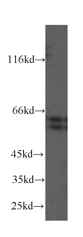
HeLa cells were subjected to SDS PAGE followed by western blot with Catalog No:107567(AKT antibody) at dilution of 1:1000
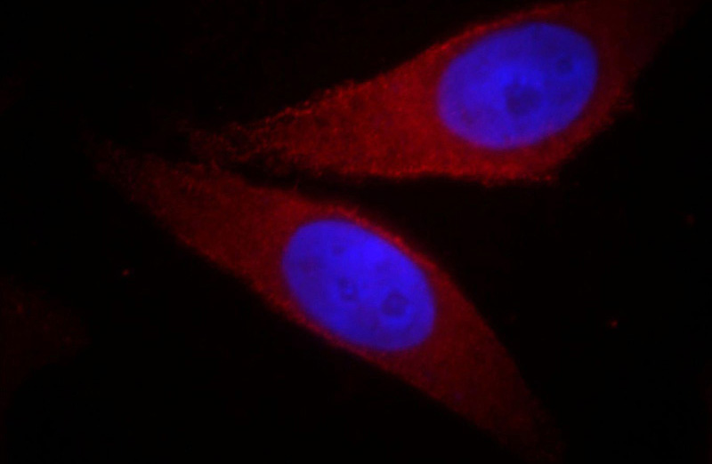
Immunofluorescent analysis of MCF-7 cells, using AKT1 antibody Catalog No: at 1:50 dilution and Rhodamine-labeled goat anti-mouse IgG (red). Blue pseudocolor = DAPI (fluorescent DNA dye).
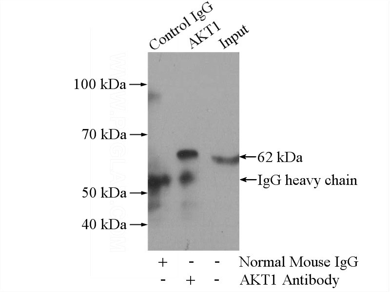
IP Result of anti-AKT (IP:Catalog No:107567, 5ug; Detection:Catalog No:107567 1:600) with mouse brain tissue lysate 3440ug.
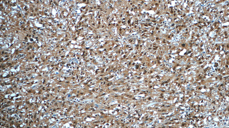
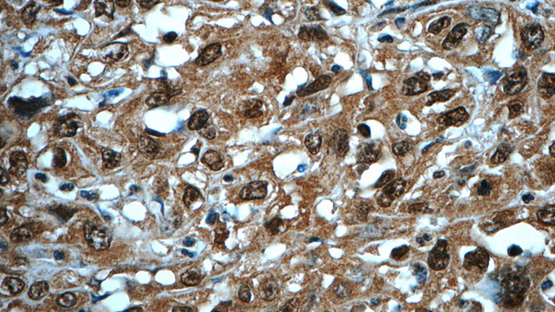
Immunohistochemistry of paraffin-embedded human cervical cancer tissue slide using Catalog No:107567(AKT Antibody) at dilution of 1:50 (under 10x lens)
Immunohistochemistry of paraffin-embedded human cervical cancer tissue slide using Catalog No:107567(AKT Antibody) at dilution of 1:50 (under 40x lens)
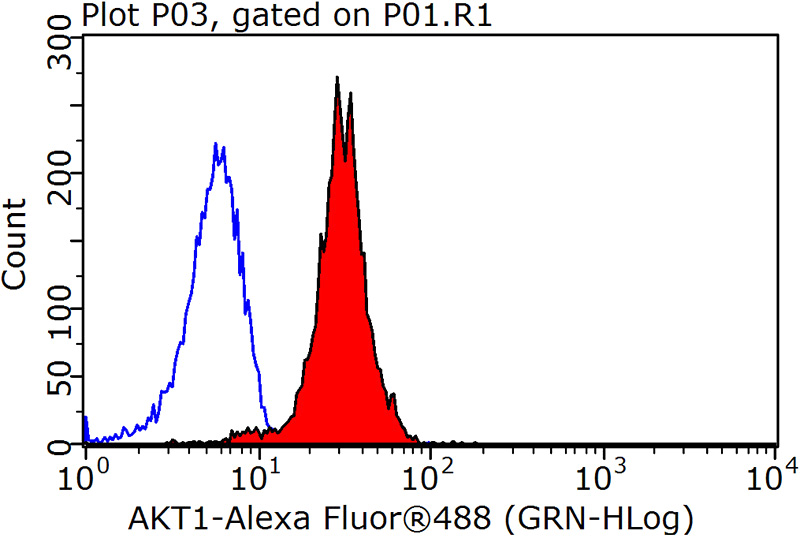
1X10^6 MCF-7 cells were stained with 0.2ug AKT antibody (Catalog No:107567, red) and control antibody (blue). Fixed with 90% MeOH blocked with 3% BSA (30 min). Alexa Fluor 488-congugated AffiniPure Goat Anti-Mouse IgG(H+L) with dilution 1:1500.
-
Background
The serine-threonine protein kinase AKT1 is catalytically inactive in serum-starved primary and immortalized fibroblasts. Survival factors can suppress apoptosis in a transcription-independent manner by activating the serine/threonine kinase AKT1, which then phosphorylates and inactivates components of the apoptotic machinery.
Related Products / Services
Please note: All products are "FOR RESEARCH USE ONLY AND ARE NOT INTENDED FOR DIAGNOSTIC OR THERAPEUTIC USE"