
-
Product Name
AKAP7 antibody
- Documents
-
Description
AKAP7 Rabbit Polyclonal antibody. Positive IHC detected in human brain tissue, human heart tissue. Positive IF detected in HepG2 cells. Positive IP detected in mouse brain tissue. Positive WB detected in human brain tissue, human heart tissue. Observed molecular weight by Western-blot: 11 kDa,18 kDa,37 kDa
-
Tested applications
ELISA, IHC, IF, WB, IP
-
Species reactivity
Human,Mouse,Rat; other species not tested.
-
Alternative names
A kinase anchor protein 18 kDa antibody; AKAP 18 antibody; AKAP 7 isoforms alpha and beta antibody; AKAP18 antibody; AKAP7 antibody
-
Isotype
Rabbit IgG
-
Preparation
This antibody was obtained by immunization of AKAP7 recombinant protein (Accession Number: NM_004842). Purification method: Antigen affinity purified.
-
Clonality
Polyclonal
-
Formulation
PBS with 0.02% sodium azide and 50% glycerol pH 7.3.
-
Storage instructions
Store at -20℃. DO NOT ALIQUOT
-
Applications
Recommended Dilution:
WB: 1:200-1:2000
IP: 1:200-1:1000
IHC: 1:20-1:200
IF: 1:10-1:100
-
Validations
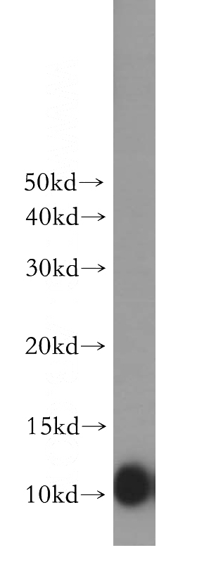
human brain tissue were subjected to SDS PAGE followed by western blot with Catalog No:107934(AKAP7 antibody) at dilution of 1:500
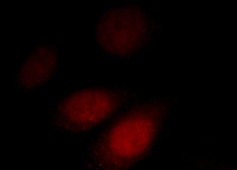
Immunofluorescent analysis of HepG2 cells, using AKAP7 antibody Catalog No:107934 at 1:25 dilution and Rhodamine-labeled goat anti-rabbit IgG (red).
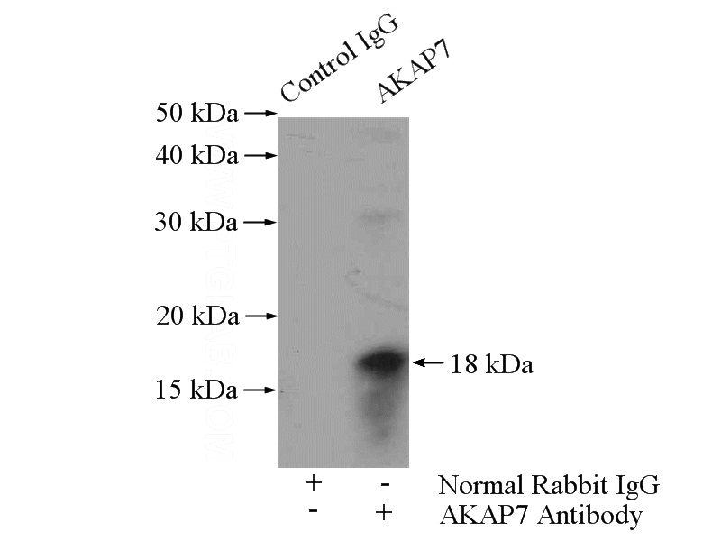
IP Result of anti-AKAP7 (IP:Catalog No:107934, 4ug; Detection:Catalog No:107934 1:300) with mouse brain tissue lysate 4000ug.
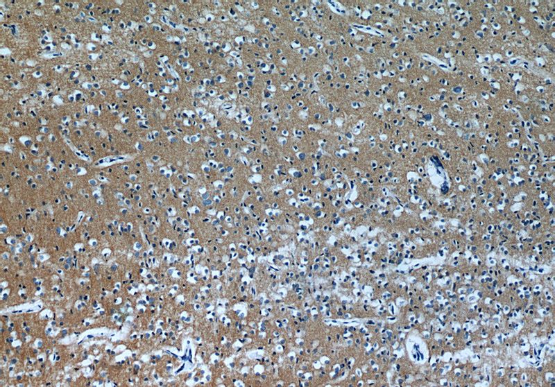
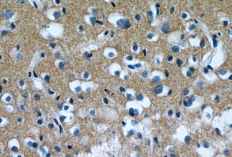
Immunohistochemistry of paraffin-embedded human brain tissue slide using Catalog No:107934(AKAP7 Antibody) at dilution of 1:50 (under 10x lens)
Immunohistochemistry of paraffin-embedded human brain tissue slide using Catalog No:107934(AKAP7 Antibody) at dilution of 1:50 (under 40x lens)
-
Background
AKAP7, also named as AKAP15 and AKAP18, localize PKA in complexes with CaV1.2 and PLN, respectively. AKAP7 targets the cAMP-dependent protein kinase (PKA) to the plasma membrane, and permits functional coupling to the L-type calcium channel. AKAP7 has short isoform AKAP7 alpha and AKAP7 beta with MW 15-18 kDa; long isoform AKAP7 gamma with MW 37-42 kDa. AKAP7alpha was highly expressed only in brain and weakly in lung lysates from WT animals. This antibody can recognize all the isoforms of AKAP7.
-
References
- Jones BW, Brunet S, Gilbert ML. Cardiomyocytes from AKAP7 knockout mice respond normally to adrenergic stimulation. Proceedings of the National Academy of Sciences of the United States of America. 109(42):17099-104. 2012.
- Gusho E, Zhang R, Jha BK. Murine AKAP7 has a 2',5'-phosphodiesterase domain that can complement an inactive murine coronavirus ns2 gene. mBio. 5(4):e01312-14. 2014.
Related Products / Services
Please note: All products are "FOR RESEARCH USE ONLY AND ARE NOT INTENDED FOR DIAGNOSTIC OR THERAPEUTIC USE"