
-
Product Name
AGR2 antibody
- Documents
-
Description
AGR2 Rabbit Polyclonal antibody. Positive WB detected in mouse stomach tissue, human colon tissue, human stomach tissue, mouse small intestine tissue, rat colon tissue, rat stomach tissue, SW 1990 cells. Positive IP detected in SW 1990 cells. Positive IHC detected in human breast cancer tissue, human colon tissue. Positive IF detected in MCF-7 cells. Positive FC detected in MCF-7 cells. Observed molecular weight by Western-blot: 17-20kd
-
Tested applications
ELISA, WB, IHC, IP, FC, IF
-
Species reactivity
Human,Mouse,Rat; other species not tested.
-
Alternative names
AG 2 antibody; AG2 antibody; AGR2 antibody; GOB 4 antibody; HAG 2 antibody; HPC8 antibody; PDIA17 antibody; XAG 2 antibody
-
Isotype
Rabbit IgG
-
Preparation
This antibody was obtained by immunization of AGR2 recombinant protein (Accession Number: XM_005249581). Purification method: Antigen affinity purified.
-
Clonality
Polyclonal
-
Formulation
PBS with 0.1% sodium azide and 50% glycerol pH 7.3.
-
Storage instructions
Store at -20℃. DO NOT ALIQUOT
-
Applications
Recommended Dilution:
WB: 1:1000-1:10000
IP: 1:500-1:5000
IHC: 1:20-1:200
IF: 1:10-1:100
-
Validations
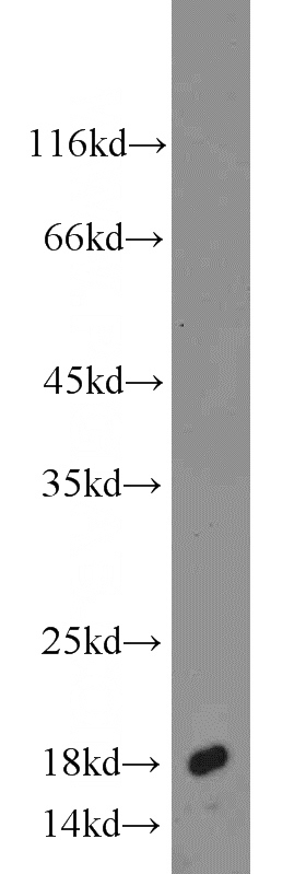
mouse stomach tissue were subjected to SDS PAGE followed by western blot with Catalog No:107835(AGR2 antibody) at dilution of 1:1000
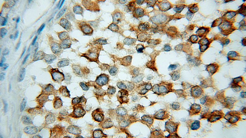
Immunohistochemical of paraffin-embedded human breast cancer using Catalog No:107835(AGR2 antibody) at dilution of 1:100 (under 10x lens)
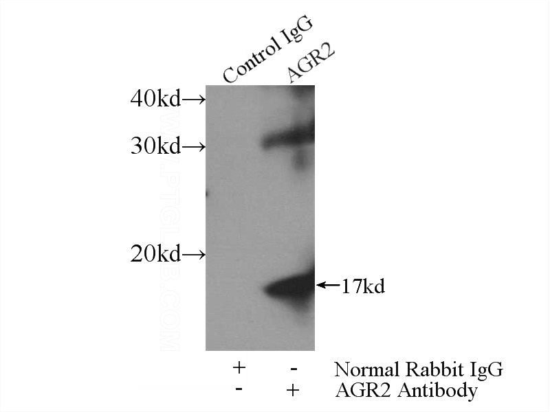
IP Result of anti-AGR2 (IP:Catalog No:107835, 4ug; Detection:Catalog No:107835 1:1000) with SW 1990 cells lysate 2400ug.
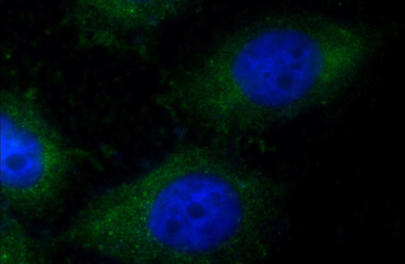
Immunofluorescent analysis of MCF-7 cells using Catalog No:107835(AGR2 Antibody) at dilution of 1:25 and Alexa Fluor 488-congugated AffiniPure Goat Anti-Rabbit IgG(H+L)
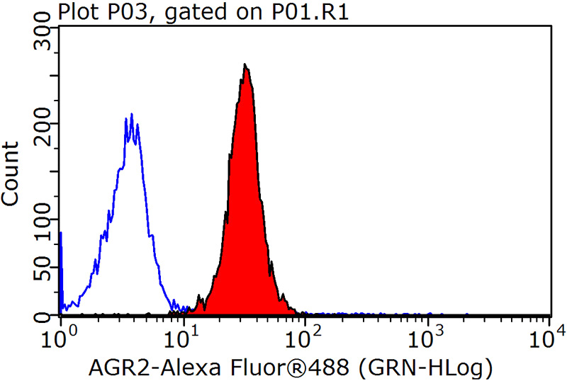
1X10^6 MCF-7 cells were stained with 0.2ug AGR2 antibody (Catalog No:107835, red) and control antibody (blue). Fixed with 90% MeOH blocked with 3% BSA (30 min). Alexa Fluor 488-congugated AffiniPure Goat Anti-Rabbit IgG(H+L) with dilution 1:1500.
-
Background
AGR2, also named AG2 or HPC8, encodes anterior gradient protein 2 homolog which belongs to the AGR family. It is a secreted protein localized in endoplasmic reticulum. AGR2 plays roles in MUC2 post-transcriptional synthesis,secretion and production of mucus by intestinal cells. AGR2 was significantly elevated in the pancreatic juice from patients with pre-malignant conditions as well as pancreatic cancer compared to control pancreatic juice samples. AGR2 levels in pancreatic juice could potentially be used to aide in assessment of high-risk patients undergoing endoscopic procedures.
-
References
- Xue H, Lü B, Zhang J. Identification of serum biomarkers for colorectal cancer metastasis using a differential secretome approach. Journal of proteome research. 9(1):545-55. 2010.
- Herfs M, Yamamoto Y, Laury A. A discrete population of squamocolumnar junction cells implicated in the pathogenesis of cervical cancer. Proceedings of the National Academy of Sciences of the United States of America. 109(26):10516-21. 2012.
- Herfs M, Vargas SO, Yamamoto Y. A novel blueprint for 'top down' differentiation defines the cervical squamocolumnar junction during development, reproductive life, and neoplasia. The Journal of pathology. 229(3):460-8. 2013.
- Herfs M, Parra-Herran C, Howitt BE. Cervical squamocolumnar junction-specific markers define distinct, clinically relevant subsets of low-grade squamous intraepithelial lesions. The American journal of surgical pathology. 37(9):1311-8. 2013.
- Herfs M, Somja J, Howitt BE. Unique recurrence patterns of cervical intraepithelial neoplasia after excision of the squamocolumnar junction. International journal of cancer. 136(5):1043-52. 2015.
Related Products / Services
Please note: All products are "FOR RESEARCH USE ONLY AND ARE NOT INTENDED FOR DIAGNOSTIC OR THERAPEUTIC USE"